

promising cellular potency. Their aminopyrimidine series comprised about 500
compounds and 12 examples, all showing no inhibition toward p38α MAP
kinase at a test concentration of 20 μM, were further profiled [24]. Compound
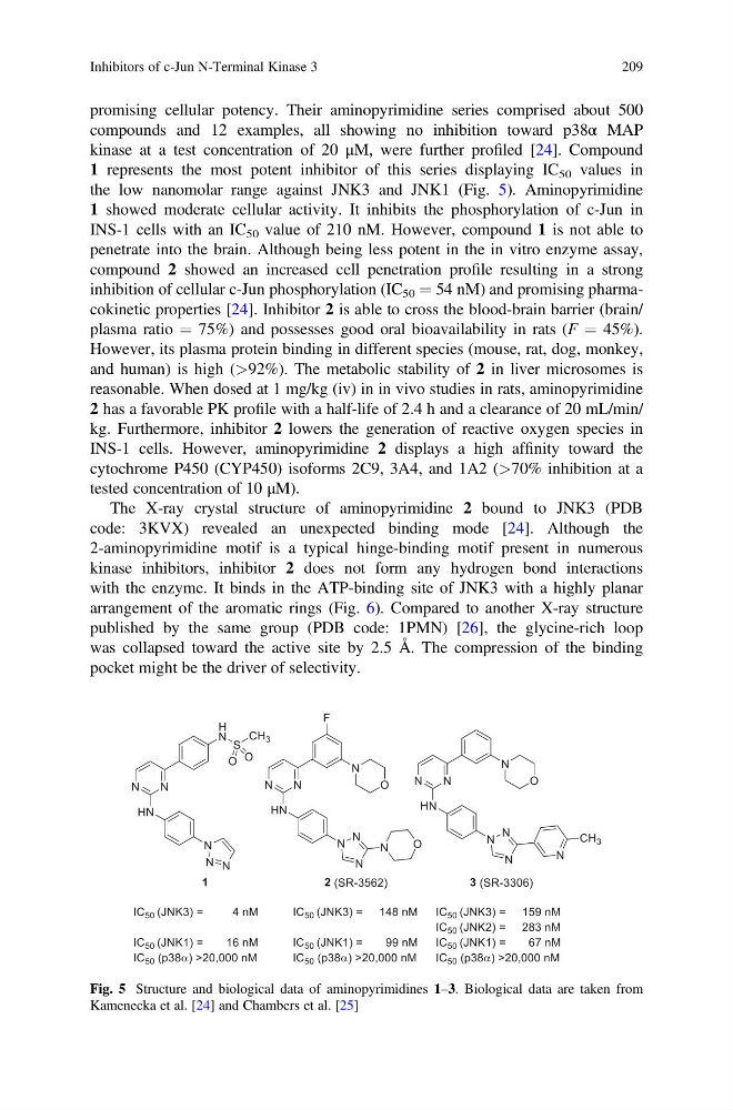
1 represents the most potent inhibitor of this series displaying IC 50 values in
the low nanomolar range against JNK3 and JNK1 (Fig. 5). Aminopyrimidine
1 showed moderate cellular activity. It inhibits the phosphorylation of c-Jun in
INS-1 cells with an IC 50 value of 210 nM. However, compound 1 is not able to
penetrate into the brain. Although being less potent in the in vitro enzyme assay,
compound 2 showed an increased cell penetration profile resulting in a strong
inhibition of cellular c-Jun phosphorylation (IC 50 ¼ 54 nM) and promising pharmacokinetic properties [24]. Inhibitor 2 is able to cross the blood-brain barrier (brain/
plasma ratio ¼ 75%) and possesses good oral bioavailability in rats (F ¼ 45%).
However, its plasma protein binding in different species (mouse, rat, dog, monkey,
and human) is high (>92%). The metabolic stability of 2 in liver microsomes is
reasonable. When dosed at 1 mg/kg (iv) in in vivo studies in rats, aminopyrimidine
2 has a favorable PK profile with a half-life of 2.4 h and a clearance of 20 mL/min/
kg. Furthermore, inhibitor 2 lowers the generation of reactive oxygen species in
INS-1 cells. However, aminopyrimidine 2 displays a high affinity toward the
cytochrome P450 (CYP450) isoforms 2C9, 3A4, and 1A2 (>70% inhibition at a
tested concentration of 10 μM).
The X-ray crystal structure of aminopyrimidine 2 bound to JNK3 (PDB
code: 3KVX) revealed an unexpected binding mode [24]. Although the
2-aminopyrimidine motif is a typical hinge-binding motif present in numerous
kinase inhibitors, inhibitor 2 does not form any hydrogen bond interactions
with the enzyme. It binds in the ATP-binding site of JNK3 with a highly planar
arrangement of the aromatic rings (Fig. 6). Compared to another X-ray structure
published by the same group (PDB code: 1PMN) [26], the glycine-rich loop
was collapsed toward the active site by 2.5 Å. The compression of the binding
pocket might be the driver of selectivity.
Fig. 5 Structure and biological data of aminopyrimidines 1–3. Biological data are taken from
Kamenecka et al. [24] and Chambers et al. [25]
Inhibitors of c-Jun N-Terminal Kinase 3
209
compounds and 12 examples, all showing no inhibition toward p38α MAP
kinase at a test concentration of 20 μM, were further profiled [24]. Compound
1 represents the most potent inhibitor of this series displaying IC 50 values in
the low nanomolar range against JNK3 and JNK1 (Fig. 5). Aminopyrimidine
1 showed moderate cellular activity. It inhibits the phosphorylation of c-Jun in
INS-1 cells with an IC 50 value of 210 nM. However, compound 1 is not able to
penetrate into the brain. Although being less potent in the in vitro enzyme assay,
compound 2 showed an increased cell penetration profile resulting in a strong
inhibition of cellular c-Jun phosphorylation (IC 50 ¼ 54 nM) and promising pharmacokinetic properties [24]. Inhibitor 2 is able to cross the blood-brain barrier (brain/
plasma ratio ¼ 75%) and possesses good oral bioavailability in rats (F ¼ 45%).
However, its plasma protein binding in different species (mouse, rat, dog, monkey,
and human) is high (>92%). The metabolic stability of 2 in liver microsomes is
reasonable. When dosed at 1 mg/kg (iv) in in vivo studies in rats, aminopyrimidine
2 has a favorable PK profile with a half-life of 2.4 h and a clearance of 20 mL/min/
kg. Furthermore, inhibitor 2 lowers the generation of reactive oxygen species in
INS-1 cells. However, aminopyrimidine 2 displays a high affinity toward the
cytochrome P450 (CYP450) isoforms 2C9, 3A4, and 1A2 (>70% inhibition at a
tested concentration of 10 μM).
The X-ray crystal structure of aminopyrimidine 2 bound to JNK3 (PDB
code: 3KVX) revealed an unexpected binding mode [24]. Although the
2-aminopyrimidine motif is a typical hinge-binding motif present in numerous
kinase inhibitors, inhibitor 2 does not form any hydrogen bond interactions
with the enzyme. It binds in the ATP-binding site of JNK3 with a highly planar
arrangement of the aromatic rings (Fig. 6). Compared to another X-ray structure
published by the same group (PDB code: 1PMN) [26], the glycine-rich loop
was collapsed toward the active site by 2.5 Å. The compression of the binding
pocket might be the driver of selectivity.
Fig. 5 Structure and biological data of aminopyrimidines 1–3. Biological data are taken from
Kamenecka et al. [24] and Chambers et al. [25]
Inhibitors of c-Jun N-Terminal Kinase 3
209