

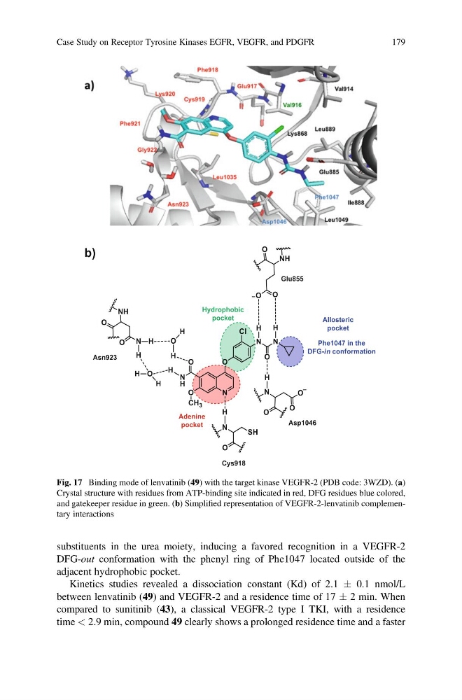
substituents in the urea moiety, inducing a favored recognition in a VEGFR-2
DFG-out conformation with the phenyl ring of Phe1047 located outside of the
adjacent hydrophobic pocket.
Kinetics studies revealed a dissociation constant (Kd) of 2.1 Æ 0.1 nmol/L
between lenvatinib (49) and VEGFR-2 and a residence time of 17 Æ 2 min. When
compared to sunitinib (43), a classical VEGFR-2 type I TKI, with a residence
time < 2.9 min, compound 49 clearly shows a prolonged residence time and a faster
Fig. 17 Binding mode of lenvatinib (49) with the target kinase VEGFR-2 (PDB code: 3WZD). (a)
Crystal structure with residues from ATP-binding site indicated in red, DFG residues blue colored,
and gatekeeper residue in green. (b) Simplified representation of VEGFR-2-lenvatinib complementary interactions
Case Study on Receptor Tyrosine Kinases EGFR, VEGFR, and PDGFR
179
DFG-out conformation with the phenyl ring of Phe1047 located outside of the
adjacent hydrophobic pocket.
Kinetics studies revealed a dissociation constant (Kd) of 2.1 Æ 0.1 nmol/L
between lenvatinib (49) and VEGFR-2 and a residence time of 17 Æ 2 min. When
compared to sunitinib (43), a classical VEGFR-2 type I TKI, with a residence
time < 2.9 min, compound 49 clearly shows a prolonged residence time and a faster
Fig. 17 Binding mode of lenvatinib (49) with the target kinase VEGFR-2 (PDB code: 3WZD). (a)
Crystal structure with residues from ATP-binding site indicated in red, DFG residues blue colored,
and gatekeeper residue in green. (b) Simplified representation of VEGFR-2-lenvatinib complementary interactions
Case Study on Receptor Tyrosine Kinases EGFR, VEGFR, and PDGFR
179