

Coarse-Grained Modeling and Simulations of Thermoresponsive …
43
the reduced temperature of T
∗
= 5.92 mimics T = 298K. Masses of all BB beads
and HB beads are m
BB
= 3m and m
HB
= 1m, respectively. The units are chosen in
such a way that the size and mass of a BB bead roughly matches that of a nucleotide;
however, these do not match the size and mass of each nucleotide precisely as this
is a generic model for varying ONA backbone chemistry. Furthermore, as it is the
interactions between CG beads rather than the mass of the CG beads that govern the
equilibrium structure and thermodynamics of the system, the choice of the CG bead
masses is inconsequential. Figure 1 shows a fully hybridized duplex represented
with our CG model. Similar to the CG DNA model of Starr and Sciortino [15],
our model does not reproduce the experimentally observed helicity of DNA as it
has been designed to capture the correct thermodynamics of DNA melting, and in
general, ONA duplex melting.
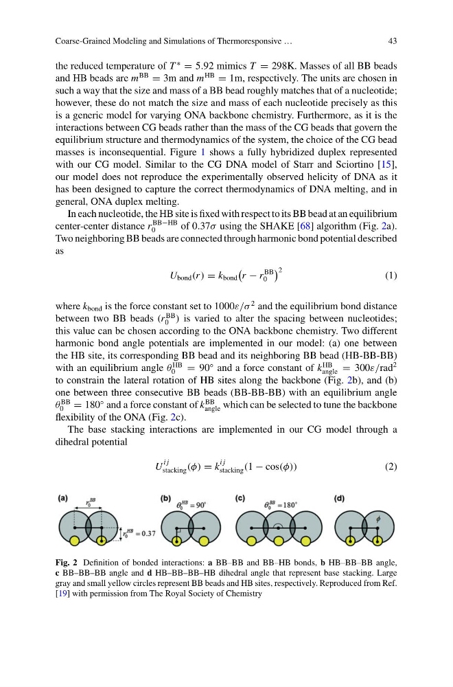
In each nucleotide, the HB site is fixed with respect to its BB bead at an equilibrium
center-center distance r
BB−HB
0
of 0.37σ using the SHAKE [68] algorithm (Fig. 2a).
Two neighboring BB beads are connected through harmonic bond potential described
as
U bond (r ) = k bond
r − r
BB
0
2
(1)
where k bond is the force constant set to 1000ε/σ
2 and the equilibrium bond distance
between two BB beads (r
BB
0 ) is varied to alter the spacing between nucleotides;
this value can be chosen according to the ONA backbone chemistry. Two different
harmonic bond angle potentials are implemented in our model: (a) one between
the HB site, its corresponding BB bead and its neighboring BB bead (HB-BB-BB)
with an equilibrium angle θ
HB
0
= 90
◦ and a force constant of k
HB
angle = 300ε/rad
2
to constrain the lateral rotation of HB sites along the backbone (Fig. 2b), and (b)
one between three consecutive BB beads (BB-BB-BB) with an equilibrium angle
θ
BB
0 = 180
◦ and a force constant of k
BB
angle which can be selected to tune the backbone
flexibility of the ONA (Fig. 2c).
The base stacking interactions are implemented in our CG model through a
dihedral potential
U
i j
stacking (φ) = k
i j
stacking (1 − cos(φ))
(2)
Fig. 2 Definition of bonded interactions: a BB–BB and BB–HB bonds, b HB–BB–BB angle,
c BB–BB–BB angle and d HB–BB–BB–HB dihedral angle that represent base stacking. Large
gray and small yellow circles represent BB beads and HB sites, respectively. Reproduced from Ref.
[19] with permission from The Royal Society of Chemistry
43
the reduced temperature of T
∗
= 5.92 mimics T = 298K. Masses of all BB beads
and HB beads are m
BB
= 3m and m
HB
= 1m, respectively. The units are chosen in
such a way that the size and mass of a BB bead roughly matches that of a nucleotide;
however, these do not match the size and mass of each nucleotide precisely as this
is a generic model for varying ONA backbone chemistry. Furthermore, as it is the
interactions between CG beads rather than the mass of the CG beads that govern the
equilibrium structure and thermodynamics of the system, the choice of the CG bead
masses is inconsequential. Figure 1 shows a fully hybridized duplex represented
with our CG model. Similar to the CG DNA model of Starr and Sciortino [15],
our model does not reproduce the experimentally observed helicity of DNA as it
has been designed to capture the correct thermodynamics of DNA melting, and in
general, ONA duplex melting.
In each nucleotide, the HB site is fixed with respect to its BB bead at an equilibrium
center-center distance r
BB−HB
0
of 0.37σ using the SHAKE [68] algorithm (Fig. 2a).
Two neighboring BB beads are connected through harmonic bond potential described
as
U bond (r ) = k bond
r − r
BB
0
2
(1)
where k bond is the force constant set to 1000ε/σ
2 and the equilibrium bond distance
between two BB beads (r
BB
0 ) is varied to alter the spacing between nucleotides;
this value can be chosen according to the ONA backbone chemistry. Two different
harmonic bond angle potentials are implemented in our model: (a) one between
the HB site, its corresponding BB bead and its neighboring BB bead (HB-BB-BB)
with an equilibrium angle θ
HB
0
= 90
◦ and a force constant of k
HB
angle = 300ε/rad
2
to constrain the lateral rotation of HB sites along the backbone (Fig. 2b), and (b)
one between three consecutive BB beads (BB-BB-BB) with an equilibrium angle
θ
BB
0 = 180
◦ and a force constant of k
BB
angle which can be selected to tune the backbone
flexibility of the ONA (Fig. 2c).
The base stacking interactions are implemented in our CG model through a
dihedral potential
U
i j
stacking (φ) = k
i j
stacking (1 − cos(φ))
(2)
Fig. 2 Definition of bonded interactions: a BB–BB and BB–HB bonds, b HB–BB–BB angle,
c BB–BB–BB angle and d HB–BB–BB–HB dihedral angle that represent base stacking. Large
gray and small yellow circles represent BB beads and HB sites, respectively. Reproduced from Ref.
[19] with permission from The Royal Society of Chemistry