

42
A. Jayaraman et al.
conjugated to solvophilic homopolymers [20, 66]. Ghobadi and Jayaraman [20]
observed that the conjugation of solvophilic homopolymer with ONAs does not
impact the melting temperature for ONA strands with high G-C content irrespective of the polymer length (8 repeat units to 100 repeat units), polymer architecture
(linear or short 4-arm star), ONA flexibility (flexible and semi-flexible ONAs) and
ONA backbone charge (electrostatically neutral and negatively charged). Prhashanna
and Jayaraman [67] extended the above exploration to ONAs conjugated to solvophobic linear homopolymers and found that the melting temperature increases for
charge neutral and flexible ONAs (similar to PNA) while it decreases for negatively
charged and semi-flexible ONAs (similar to DNA) upon conjugation with relatively
solvophobic long homopolymer (40 repeat units to 100 repeat units) for 8-mer ONAs.
In the following sections, we present the details of this h-bonding CG model for
ONAs and ONA-polymer conjugates developed in our research group [19, 20, 66,
67] which has the ability to reproduce experimentally observed melting curves of
DNA and physically expected trends in the melting profile of other ONAs.
2.2 Model
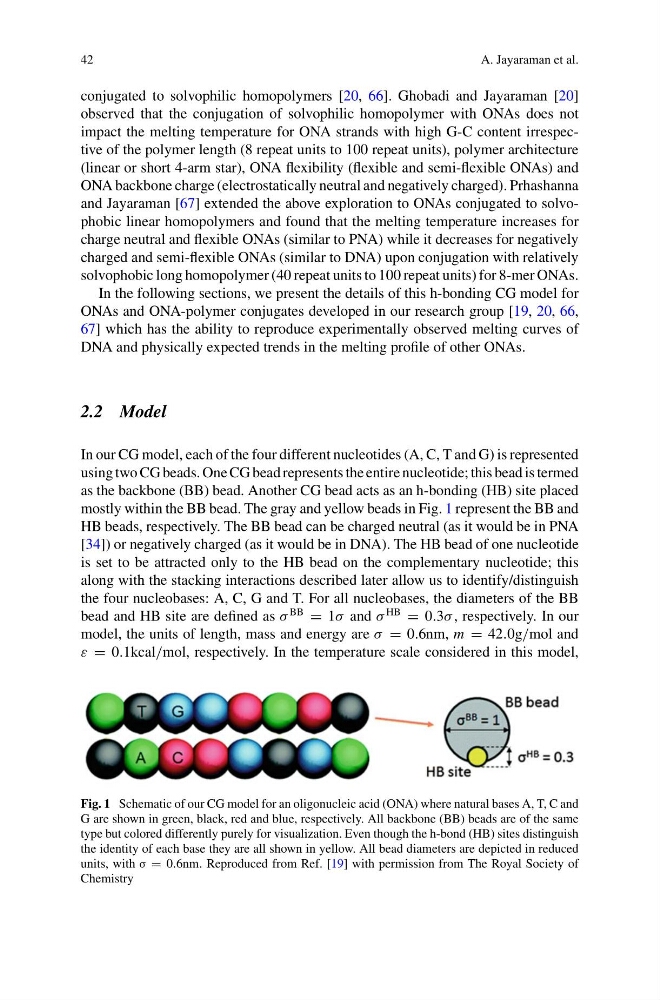
In our CG model, each of the four different nucleotides (A, C, T and G) is represented
using two CG beads. One CG bead represents the entire nucleotide; this bead is termed
as the backbone (BB) bead. Another CG bead acts as an h-bonding (HB) site placed
mostly within the BB bead. The gray and yellow beads in Fig. 1 represent the BB and
HB beads, respectively. The BB bead can be charged neutral (as it would be in PNA
[34]) or negatively charged (as it would be in DNA). The HB bead of one nucleotide
is set to be attracted only to the HB bead on the complementary nucleotide; this
along with the stacking interactions described later allow us to identify/distinguish
the four nucleobases: A, C, G and T. For all nucleobases, the diameters of the BB
bead and HB site are defined as σ
BB
= 1σ and σ
HB
= 0.3σ , respectively. In our
model, the units of length, mass and energy are σ = 0.6nm, m = 42.0g/mol and
ε = 0.1kcal/mol, respectively. In the temperature scale considered in this model,
Fig. 1 Schematic of our CG model for an oligonucleic acid (ONA) where natural bases A, T, C and
G are shown in green, black, red and blue, respectively. All backbone (BB) beads are of the same
type but colored differently purely for visualization. Even though the h-bond (HB) sites distinguish
the identity of each base they are all shown in yellow. All bead diameters are depicted in reduced
units, with σ = 0.6nm. Reproduced from Ref. [19] with permission from The Royal Society of
Chemistry
A. Jayaraman et al.
conjugated to solvophilic homopolymers [20, 66]. Ghobadi and Jayaraman [20]
observed that the conjugation of solvophilic homopolymer with ONAs does not
impact the melting temperature for ONA strands with high G-C content irrespective of the polymer length (8 repeat units to 100 repeat units), polymer architecture
(linear or short 4-arm star), ONA flexibility (flexible and semi-flexible ONAs) and
ONA backbone charge (electrostatically neutral and negatively charged). Prhashanna
and Jayaraman [67] extended the above exploration to ONAs conjugated to solvophobic linear homopolymers and found that the melting temperature increases for
charge neutral and flexible ONAs (similar to PNA) while it decreases for negatively
charged and semi-flexible ONAs (similar to DNA) upon conjugation with relatively
solvophobic long homopolymer (40 repeat units to 100 repeat units) for 8-mer ONAs.
In the following sections, we present the details of this h-bonding CG model for
ONAs and ONA-polymer conjugates developed in our research group [19, 20, 66,
67] which has the ability to reproduce experimentally observed melting curves of
DNA and physically expected trends in the melting profile of other ONAs.
2.2 Model
In our CG model, each of the four different nucleotides (A, C, T and G) is represented
using two CG beads. One CG bead represents the entire nucleotide; this bead is termed
as the backbone (BB) bead. Another CG bead acts as an h-bonding (HB) site placed
mostly within the BB bead. The gray and yellow beads in Fig. 1 represent the BB and
HB beads, respectively. The BB bead can be charged neutral (as it would be in PNA
[34]) or negatively charged (as it would be in DNA). The HB bead of one nucleotide
is set to be attracted only to the HB bead on the complementary nucleotide; this
along with the stacking interactions described later allow us to identify/distinguish
the four nucleobases: A, C, G and T. For all nucleobases, the diameters of the BB
bead and HB site are defined as σ
BB
= 1σ and σ
HB
= 0.3σ , respectively. In our
model, the units of length, mass and energy are σ = 0.6nm, m = 42.0g/mol and
ε = 0.1kcal/mol, respectively. In the temperature scale considered in this model,
Fig. 1 Schematic of our CG model for an oligonucleic acid (ONA) where natural bases A, T, C and
G are shown in green, black, red and blue, respectively. All backbone (BB) beads are of the same
type but colored differently purely for visualization. Even though the h-bond (HB) sites distinguish
the identity of each base they are all shown in yellow. All bead diameters are depicted in reduced
units, with σ = 0.6nm. Reproduced from Ref. [19] with permission from The Royal Society of
Chemistry