

9 Analytical Protocols in Antioxidant Capacity Measurement
209
for the same tetrapirolic structures, probably due to the physical structure of the test
system, the nature of the oxidation substrate, and the analytical method employed
(Becker et al. 2004).
The mechanism of antioxidant action of chlorophylls is not yet well understood;
however, the conclusions about proportionality between the CBDs system and the
ability to scavenge radicals established in the literature suggest that chlorophyll acts
as an effective electron donor (see Fig. 9.2) (Kumar et al. 2001; Lanfer-Marquez
et al. 2005; Fernandes et al. 2017). Despite being the most abundant structure in
most studies, chlorophylls were not included in the trials (Lanfer-Marquez et al.
2005). As far as our knowledge is concerned, Rodrigues et al. (2015) and Fernandes
et al. (2017) were the only ones to evaluate the antioxidant capacity of a microalgae
chlorophyll extract. Rodrigues et al (2015) demonstrated that chlorophyll extract of
Phormidium autumnale microalgae was almost 85 times more efficient as peroxyl
radical scavenger than α-tocopherol in lipophilic media. Furthermore, in their findings, Fernandes et al. (2017) showed that commercial standards of chlorophylls a
and b have higher ability to scavenge peroxyl radicals than P. autumnale extract with
11 characterized structures including chlorophyll a, b, and its derivatives.
In addition to these typical pigments, phycobiliproteins, which are open tetrapyrrole accessory pigments, they are also produced by aquatic microorganisms (Chen
and Blankenship 2011). The main phycobiliproteins produced and marketed are
phycoerythrin and phycocyanin from cyanobacteria and Rhodophyta (Viskari and
Colyer 2003). In addition, these compounds are also found in a class of biflagellate
unicellular eukaryotic algae (cryptomonads) (Román et al. 2002).
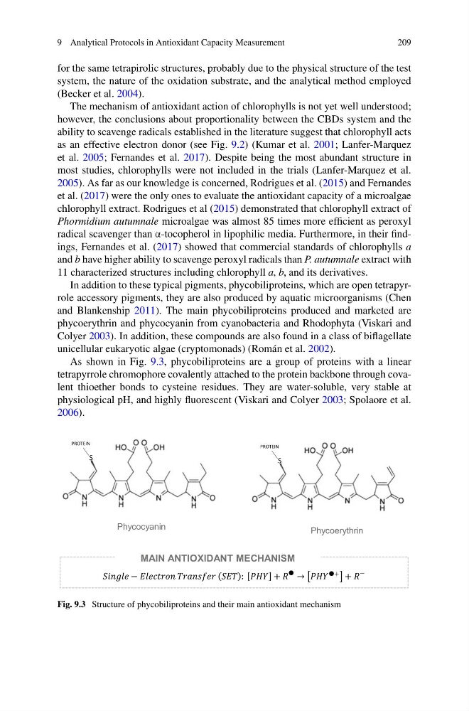
As shown in Fig. 9.3, phycobiliproteins are a group of proteins with a linear
tetrapyrrole chromophore covalently attached to the protein backbone through covalent thioether bonds to cysteine residues. They are water-soluble, very stable at
physiological pH, and highly fluorescent (Viskari and Colyer 2003; Spolaore et al.
2006).
Phycocyanin
Phycoerythrin
S
PROTEIN
S
PROTEIN
MAIN ANTIOXIDANT MECHANISM
Fig. 9.3 Structure of phycobiliproteins and their main antioxidant mechanism
209
for the same tetrapirolic structures, probably due to the physical structure of the test
system, the nature of the oxidation substrate, and the analytical method employed
(Becker et al. 2004).
The mechanism of antioxidant action of chlorophylls is not yet well understood;
however, the conclusions about proportionality between the CBDs system and the
ability to scavenge radicals established in the literature suggest that chlorophyll acts
as an effective electron donor (see Fig. 9.2) (Kumar et al. 2001; Lanfer-Marquez
et al. 2005; Fernandes et al. 2017). Despite being the most abundant structure in
most studies, chlorophylls were not included in the trials (Lanfer-Marquez et al.
2005). As far as our knowledge is concerned, Rodrigues et al. (2015) and Fernandes
et al. (2017) were the only ones to evaluate the antioxidant capacity of a microalgae
chlorophyll extract. Rodrigues et al (2015) demonstrated that chlorophyll extract of
Phormidium autumnale microalgae was almost 85 times more efficient as peroxyl
radical scavenger than α-tocopherol in lipophilic media. Furthermore, in their findings, Fernandes et al. (2017) showed that commercial standards of chlorophylls a
and b have higher ability to scavenge peroxyl radicals than P. autumnale extract with
11 characterized structures including chlorophyll a, b, and its derivatives.
In addition to these typical pigments, phycobiliproteins, which are open tetrapyrrole accessory pigments, they are also produced by aquatic microorganisms (Chen
and Blankenship 2011). The main phycobiliproteins produced and marketed are
phycoerythrin and phycocyanin from cyanobacteria and Rhodophyta (Viskari and
Colyer 2003). In addition, these compounds are also found in a class of biflagellate
unicellular eukaryotic algae (cryptomonads) (Román et al. 2002).
As shown in Fig. 9.3, phycobiliproteins are a group of proteins with a linear
tetrapyrrole chromophore covalently attached to the protein backbone through covalent thioether bonds to cysteine residues. They are water-soluble, very stable at
physiological pH, and highly fluorescent (Viskari and Colyer 2003; Spolaore et al.
2006).
Phycocyanin
Phycoerythrin
S
PROTEIN
S
PROTEIN
MAIN ANTIOXIDANT MECHANISM
Fig. 9.3 Structure of phycobiliproteins and their main antioxidant mechanism