

208
T. Casagrande do Nascimento et al.
H
COOCH 3
H
COOCH 3
D
R1
R2
R3
CH 3
CH 3
CH 3
COH
H 3 -C=H 2
COH
H 3 -C=H 2
CH 3
CH 3
CH 3
COH
CH 3
CH 3
COOCH 3
R4
R6
R5
CH 3 -CH 3
H 3 -C=H 2
H 3 -C=H 2
H 3 -C=H 2
CH 3 CH 2 COOH
CH 3 CH 2 COOH
CH 3 CH 2 COOH
R1
R2
R3
R4
R5
R6
Chlorophyll a
Chlorophyll b
Chlorophyll d
Chlorophyll f
Chlorophyll c 1
Chlorophyll c 2
Chlorophyll c 3
MAIN ANTIOXIDANT MECHANISM
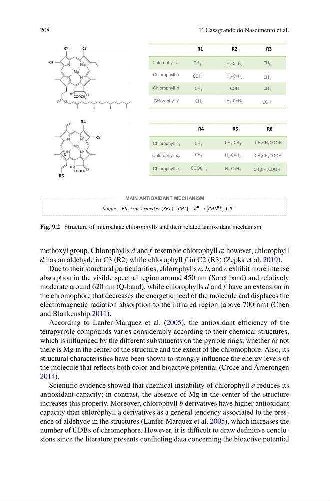
Fig. 9.2 Structure of microalgae chlorophylls and their related antioxidant mechanism
methoxyl group. Chlorophylls d and f resemble chlorophyll a; however, chlorophyll
d has an aldehyde in C3 (R2) while chlorophyll f in C2 (R3) (Zepka et al. 2019).
Due to their structural particularities, chlorophylls a, b, and c exhibit more intense
absorption in the visible spectral region around 450 nm (Soret band) and relatively
moderate around 620 nm (Q-band), while chlorophylls d and f have an extension in
the chromophore that decreases the energetic need of the molecule and displaces the
electromagnetic radiation absorption to the infrared region (above 700 nm) (Chen
and Blankenship 2011).
According to Lanfer-Marquez et al. (2005), the antioxidant efficiency of the
tetrapyrrole compounds varies considerably according to their chemical structures,
which is influenced by the different substituents on the pyrrole rings, whether or not
there is Mg in the center of the structure and the extent of the chromophore. Also, its
structural characteristics have been shown to strongly influence the energy levels of
the molecule that reflects both color and bioactive potential (Croce and Amerongen
2014).
Scientific evidence showed that chemical instability of chlorophyll a reduces its
antioxidant capacity; in contrast, the absence of Mg in the center of the structure
increases this property. Moreover, chlorophyll b derivatives have higher antioxidant
capacity than chlorophyll a derivatives as a general tendency associated to the presence of aldehyde in the structures (Lanfer-Marquez et al. 2005), which increases the
number of CDBs of chromophore. However, it is difficult to draw definitive conclusions since the literature presents conflicting data concerning the bioactive potential
T. Casagrande do Nascimento et al.
H
COOCH 3
H
COOCH 3
D
R1
R2
R3
CH 3
CH 3
CH 3
COH
H 3 -C=H 2
COH
H 3 -C=H 2
CH 3
CH 3
CH 3
COH
CH 3
CH 3
COOCH 3
R4
R6
R5
CH 3 -CH 3
H 3 -C=H 2
H 3 -C=H 2
H 3 -C=H 2
CH 3 CH 2 COOH
CH 3 CH 2 COOH
CH 3 CH 2 COOH
R1
R2
R3
R4
R5
R6
Chlorophyll a
Chlorophyll b
Chlorophyll d
Chlorophyll f
Chlorophyll c 1
Chlorophyll c 2
Chlorophyll c 3
MAIN ANTIOXIDANT MECHANISM
Fig. 9.2 Structure of microalgae chlorophylls and their related antioxidant mechanism
methoxyl group. Chlorophylls d and f resemble chlorophyll a; however, chlorophyll
d has an aldehyde in C3 (R2) while chlorophyll f in C2 (R3) (Zepka et al. 2019).
Due to their structural particularities, chlorophylls a, b, and c exhibit more intense
absorption in the visible spectral region around 450 nm (Soret band) and relatively
moderate around 620 nm (Q-band), while chlorophylls d and f have an extension in
the chromophore that decreases the energetic need of the molecule and displaces the
electromagnetic radiation absorption to the infrared region (above 700 nm) (Chen
and Blankenship 2011).
According to Lanfer-Marquez et al. (2005), the antioxidant efficiency of the
tetrapyrrole compounds varies considerably according to their chemical structures,
which is influenced by the different substituents on the pyrrole rings, whether or not
there is Mg in the center of the structure and the extent of the chromophore. Also, its
structural characteristics have been shown to strongly influence the energy levels of
the molecule that reflects both color and bioactive potential (Croce and Amerongen
2014).
Scientific evidence showed that chemical instability of chlorophyll a reduces its
antioxidant capacity; in contrast, the absence of Mg in the center of the structure
increases this property. Moreover, chlorophyll b derivatives have higher antioxidant
capacity than chlorophyll a derivatives as a general tendency associated to the presence of aldehyde in the structures (Lanfer-Marquez et al. 2005), which increases the
number of CDBs of chromophore. However, it is difficult to draw definitive conclusions since the literature presents conflicting data concerning the bioactive potential