

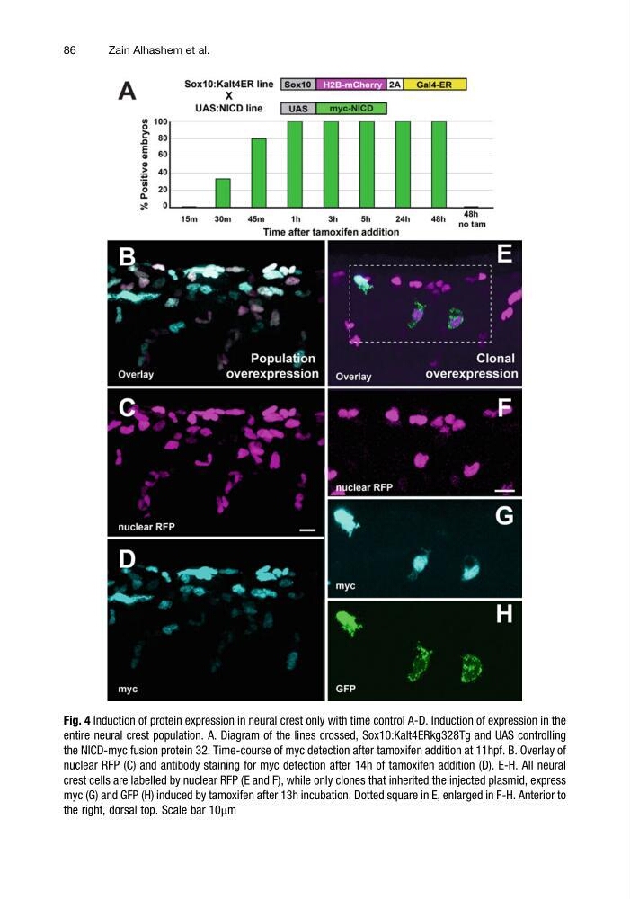
Fig. 4 Induction of protein expression in neural crest only with time control A-D. Induction of expression in the
entire neural crest population. A. Diagram of the lines crossed, Sox10:Kalt4ERkg328Tg and UAS controlling
the NICD-myc fusion protein 32. Time-course of myc detection after tamoxifen addition at 11hpf. B. Overlay of
nuclear RFP (C) and antibody staining for myc detection after 14h of tamoxifen addition (D). E-H. All neural
crest cells are labelled by nuclear RFP (E and F), while only clones that inherited the injected plasmid, express
myc (G) and GFP (H) induced by tamoxifen after 13h incubation. Dotted square in E, enlarged in F-H. Anterior to
the right, dorsal top. Scale bar 10μm
86
Zain Alhashem et al.
entire neural crest population. A. Diagram of the lines crossed, Sox10:Kalt4ERkg328Tg and UAS controlling
the NICD-myc fusion protein 32. Time-course of myc detection after tamoxifen addition at 11hpf. B. Overlay of
nuclear RFP (C) and antibody staining for myc detection after 14h of tamoxifen addition (D). E-H. All neural
crest cells are labelled by nuclear RFP (E and F), while only clones that inherited the injected plasmid, express
myc (G) and GFP (H) induced by tamoxifen after 13h incubation. Dotted square in E, enlarged in F-H. Anterior to
the right, dorsal top. Scale bar 10μm
86
Zain Alhashem et al.