

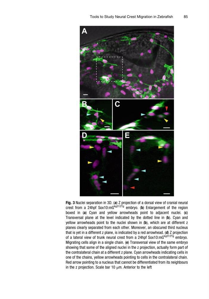
Fig. 3 Nuclei separation in 3D. (a) Z projection of a dorsal view of cranial neural
crest from a 24hpf Sox10:mG
kg312Tg embryo. (b) Enlargement of the region
boxed in (a) Cyan and yellow arrowheads point to adjacent nuclei. (c)
Transversal plane at the level indicated by the dotted line in (b). Cyan and
yellow arrowheads point to the nuclei shown in (b), which are at different z
planes clearly separated from each other. Moreover, an obscured third nucleus
that is yet in a different z plane, is indicated by a red arrowhead. (d) Z projection
of a lateral view of trunk neural crest from a 24hpf Sox10:mG
kg312Tg embryo.
Migrating cells align in a single chain. (e) Transversal view of the same embryo
showing that some of the aligned nuclei in the z projection, actually form part of
the contralateral chain at a different z plane. Cyan arrowheads indicating cells in
one of the chains, yellow arrowheads pointing to cells in the contralateral chain.
Red arrow pointing to a nucleus that cannot be differentiated from its neighbours
in the z projection. Scale bar 10 μm. Anterior to the left
Tools to Study Neural Crest Migration in Zebrafish
85
crest from a 24hpf Sox10:mG
kg312Tg embryo. (b) Enlargement of the region
boxed in (a) Cyan and yellow arrowheads point to adjacent nuclei. (c)
Transversal plane at the level indicated by the dotted line in (b). Cyan and
yellow arrowheads point to the nuclei shown in (b), which are at different z
planes clearly separated from each other. Moreover, an obscured third nucleus
that is yet in a different z plane, is indicated by a red arrowhead. (d) Z projection
of a lateral view of trunk neural crest from a 24hpf Sox10:mG
kg312Tg embryo.
Migrating cells align in a single chain. (e) Transversal view of the same embryo
showing that some of the aligned nuclei in the z projection, actually form part of
the contralateral chain at a different z plane. Cyan arrowheads indicating cells in
one of the chains, yellow arrowheads pointing to cells in the contralateral chain.
Red arrow pointing to a nucleus that cannot be differentiated from its neighbours
in the z projection. Scale bar 10 μm. Anterior to the left
Tools to Study Neural Crest Migration in Zebrafish
85