

calculate path distances, which are largely underestimated when
tracking in 2D as the movement in z is disregarded. Together,
this imaging setup and analysis tools permit the study of NC
migration in wild type conditions. The next step is to define the
molecular pathways regulating this process. General signalling perturbations such as mRNA injections or analysis of mutant embryos
can be misleading, as these manipulations may generate alterations
of the NC population early in development and/or induce defects
in other tissues. Hence, any observed NC migratory phenotypes
under such conditions may only be the result of indirect perturbations. Appropriate assessment of the signals regulating NC migration requires the analysis of gain- and loss-of-function conditions in
a tissue-specific and time-controlled manner. To attain this goal, we
have taken advantage of the UAS/Gal4 system and generated a new
Sox10:Kalt4ER
kg328Tg line (Fig. 4). The UAS system has been
extensively used in drosophila [9] and shown to work in zebrafish
[10, 11]. In the Sox10:Kalt4ER
kg328Tg transgenic, all NC have
their chromatin labelled with RFP and express the transcriptional
activator Gal4 fused to the hormone-binding domain of the oestrogen receptor (ER
T2
). In the absence of oestrogen, Gal4ER
T2 is
retained inactive in the cytoplasm. Upon addition of the estrogen
analogue tamoxifen, Gal4ER
T2 is translocated to the nucleus where
it binds and activates expression of any UAS-driven transgene. This
system can induce protein overexpression within 30 min of
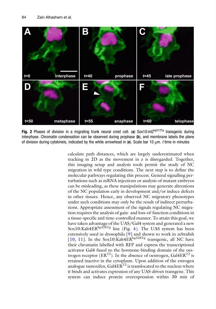
Fig. 2 Phases of division in a migrating trunk neural crest cell. (a) Sox10:mG
kg312Tg transgenic during
interphase. Chromatin condensation can be observed during prophase (b), and membrane labels the plane
of division during cytokineis, indicated by the white arrowhead in (e). Scale bar 10 μm. t time in minutes
84
Zain Alhashem et al.
tracking in 2D as the movement in z is disregarded. Together,
this imaging setup and analysis tools permit the study of NC
migration in wild type conditions. The next step is to define the
molecular pathways regulating this process. General signalling perturbations such as mRNA injections or analysis of mutant embryos
can be misleading, as these manipulations may generate alterations
of the NC population early in development and/or induce defects
in other tissues. Hence, any observed NC migratory phenotypes
under such conditions may only be the result of indirect perturbations. Appropriate assessment of the signals regulating NC migration requires the analysis of gain- and loss-of-function conditions in
a tissue-specific and time-controlled manner. To attain this goal, we
have taken advantage of the UAS/Gal4 system and generated a new
Sox10:Kalt4ER
kg328Tg line (Fig. 4). The UAS system has been
extensively used in drosophila [9] and shown to work in zebrafish
[10, 11]. In the Sox10:Kalt4ER
kg328Tg transgenic, all NC have
their chromatin labelled with RFP and express the transcriptional
activator Gal4 fused to the hormone-binding domain of the oestrogen receptor (ER
T2
). In the absence of oestrogen, Gal4ER
T2 is
retained inactive in the cytoplasm. Upon addition of the estrogen
analogue tamoxifen, Gal4ER
T2 is translocated to the nucleus where
it binds and activates expression of any UAS-driven transgene. This
system can induce protein overexpression within 30 min of
Fig. 2 Phases of division in a migrating trunk neural crest cell. (a) Sox10:mG
kg312Tg transgenic during
interphase. Chromatin condensation can be observed during prophase (b), and membrane labels the plane
of division during cytokineis, indicated by the white arrowhead in (e). Scale bar 10 μm. t time in minutes
84
Zain Alhashem et al.