

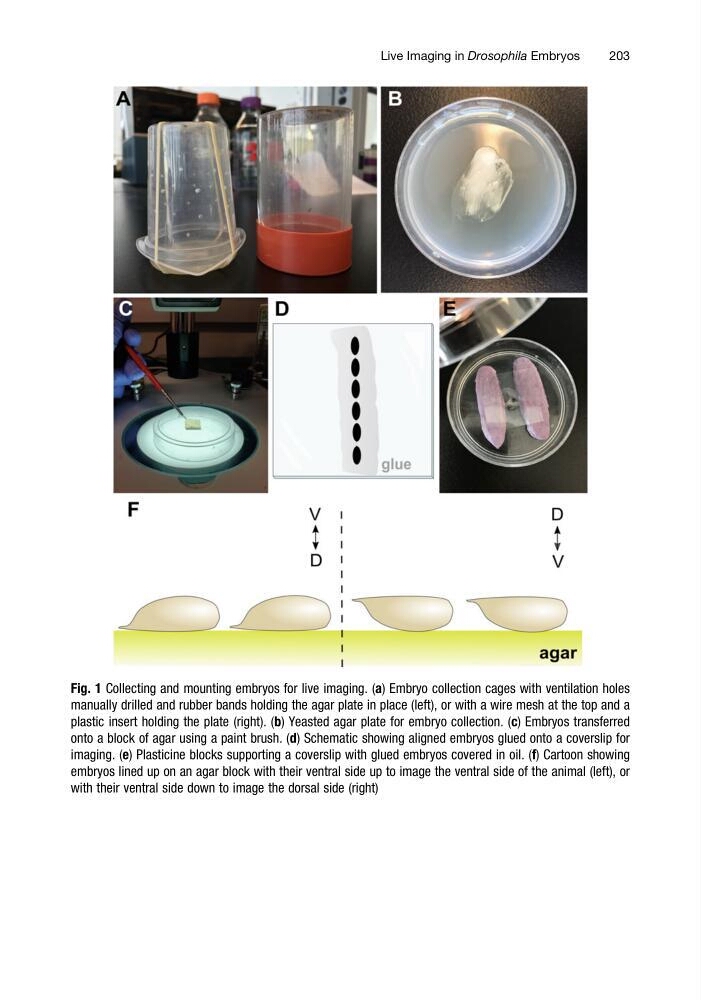
Fig. 1 Collecting and mounting embryos for live imaging. (a) Embryo collection cages with ventilation holes
manually drilled and rubber bands holding the agar plate in place (left), or with a wire mesh at the top and a
plastic insert holding the plate (right). (b) Yeasted agar plate for embryo collection. (c) Embryos transferred
onto a block of agar using a paint brush. (d) Schematic showing aligned embryos glued onto a coverslip for
imaging. (e) Plasticine blocks supporting a coverslip with glued embryos covered in oil. (f) Cartoon showing
embryos lined up on an agar block with their ventral side up to image the ventral side of the animal (left), or
with their ventral side down to image the dorsal side (right)
Live Imaging in Drosophila Embryos
203
manually drilled and rubber bands holding the agar plate in place (left), or with a wire mesh at the top and a
plastic insert holding the plate (right). (b) Yeasted agar plate for embryo collection. (c) Embryos transferred
onto a block of agar using a paint brush. (d) Schematic showing aligned embryos glued onto a coverslip for
imaging. (e) Plasticine blocks supporting a coverslip with glued embryos covered in oil. (f) Cartoon showing
embryos lined up on an agar block with their ventral side up to image the ventral side of the animal (left), or
with their ventral side down to image the dorsal side (right)
Live Imaging in Drosophila Embryos
203