

2 Materials
2.1 Embryo Staging
and Collection
1. Stereomicroscope.
2. Embryo collection cages (VWR, #25384-152 or equivalent). A
fine pin can be used to drill ventilation holes around the cage
(Fig. 1a, left). A commercial solution with a fine mesh for
ventilation at the top of the cage is also available (Diamed,
#GEN59-100 or equivalent; Fig. 1a, right).
3. 60 mm Petri dishes for embryo collection (Fig. 1b).
4. Medium to fill embryo collection plates (makes approximately
100 plates) (see Note 1): 20.25 g agar powder, 675 mL tap
water, 22.5 g sucrose, 1.35 g nipagin, and 225 mL apple juice.
5. Yeast and tap water.
6. A spatula to mix the yeast paste.
7. 50 mL tubes.
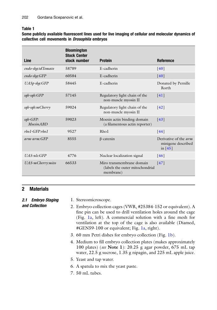
Table 1
Some publicly available fluorescent lines used for live imaging of cellular and molecular dynamics of
collective cell movements in Drosophila embryos
Line
Bloomington
Stock Center
stock number
Protein
Reference
endo-shg:tdTomato
58789
E-cadherin
[40]
endo-shg:GFP
60584
E-cadherin
[40]
UASp-shg:GFP
58445
E-cadherin
Donated by Pernille
Rorth
sqh-sqh:GFP
57145
Regulatory light chain of the
non-muscle myosin II
[41]
sqh-sqh:mCherry
59024
Regulatory light chain of the
non-muscle myosin II
[42]
sqh-GFP:
MoesinABD
59023
Moesin actin binding domain
(a filamentous actin reporter)
[43]
rho1-GFP:rho1
9527
Rho1
[44]
arm-arm:GFP
8555
β-catenin
Derivative of the arm
minigene described
in [45]
UAS-nls:GFP
4776
Nuclear localization signal
[46]
UAS-mCherry:mito
66533
Miro transmembrane domain
(labels the outer mitochondrial
membrane)
[47]
202
Gordana Scepanovic et al.
2.1 Embryo Staging
and Collection
1. Stereomicroscope.
2. Embryo collection cages (VWR, #25384-152 or equivalent). A
fine pin can be used to drill ventilation holes around the cage
(Fig. 1a, left). A commercial solution with a fine mesh for
ventilation at the top of the cage is also available (Diamed,
#GEN59-100 or equivalent; Fig. 1a, right).
3. 60 mm Petri dishes for embryo collection (Fig. 1b).
4. Medium to fill embryo collection plates (makes approximately
100 plates) (see Note 1): 20.25 g agar powder, 675 mL tap
water, 22.5 g sucrose, 1.35 g nipagin, and 225 mL apple juice.
5. Yeast and tap water.
6. A spatula to mix the yeast paste.
7. 50 mL tubes.
Table 1
Some publicly available fluorescent lines used for live imaging of cellular and molecular dynamics of
collective cell movements in Drosophila embryos
Line
Bloomington
Stock Center
stock number
Protein
Reference
endo-shg:tdTomato
58789
E-cadherin
[40]
endo-shg:GFP
60584
E-cadherin
[40]
UASp-shg:GFP
58445
E-cadherin
Donated by Pernille
Rorth
sqh-sqh:GFP
57145
Regulatory light chain of the
non-muscle myosin II
[41]
sqh-sqh:mCherry
59024
Regulatory light chain of the
non-muscle myosin II
[42]
sqh-GFP:
MoesinABD
59023
Moesin actin binding domain
(a filamentous actin reporter)
[43]
rho1-GFP:rho1
9527
Rho1
[44]
arm-arm:GFP
8555
β-catenin
Derivative of the arm
minigene described
in [45]
UAS-nls:GFP
4776
Nuclear localization signal
[46]
UAS-mCherry:mito
66533
Miro transmembrane domain
(labels the outer mitochondrial
membrane)
[47]
202
Gordana Scepanovic et al.