

plates containing Agar/Albumen culture media (Fig. 3c). Finally,
the embryos are placed in a culture chamber (Fig. 3d, e) and
examined under an upright wide-field microscope. The system we
describe here allows six specimens to be visualized simultaneously
and they can be analyzed over long periods of time, these embryos
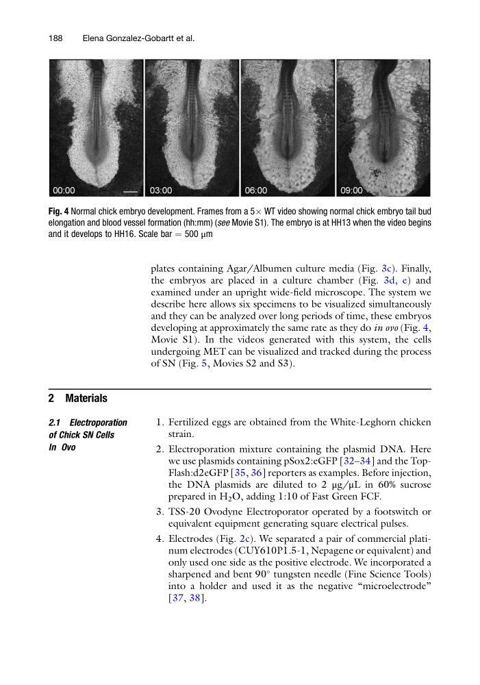
developing at approximately the same rate as they do in ovo (Fig. 4,
Movie S1). In the videos generated with this system, the cells
undergoing MET can be visualized and tracked during the process
of SN (Fig. 5, Movies S2 and S3).
2 Materials
2.1 Electroporation
of Chick SN Cells
In Ovo
1. Fertilized eggs are obtained from the White-Leghorn chicken
strain.
2. Electroporation mixture containing the plasmid DNA. Here
we use plasmids containing pSox2:eGFP [32–34] and the TopFlash:d2eGFP [35, 36] reporters as examples. Before injection,
the DNA plasmids are diluted to 2 μg/μL in 60% sucrose
prepared in H 2 O, adding 1:10 of Fast Green FCF.
3. TSS-20 Ovodyne Electroporator operated by a footswitch or
equivalent equipment generating square electrical pulses.
4. Electrodes (Fig. 2c). We separated a pair of commercial platinum electrodes (CUY610P1.5-1, Nepagene or equivalent) and
only used one side as the positive electrode. We incorporated a
sharpened and bent 90
tungsten needle (Fine Science Tools)
into a holder and used it as the negative “microelectrode”
[37, 38].
Fig. 4 Normal chick embryo development. Frames from a 5Â WT video showing normal chick embryo tail bud
elongation and blood vessel formation (hh:mm) (see Movie S1). The embryo is at HH13 when the video begins
and it develops to HH16. Scale bar ¼ 500 μm
188
Elena Gonzalez-Gobartt et al.
the embryos are placed in a culture chamber (Fig. 3d, e) and
examined under an upright wide-field microscope. The system we
describe here allows six specimens to be visualized simultaneously
and they can be analyzed over long periods of time, these embryos
developing at approximately the same rate as they do in ovo (Fig. 4,
Movie S1). In the videos generated with this system, the cells
undergoing MET can be visualized and tracked during the process
of SN (Fig. 5, Movies S2 and S3).
2 Materials
2.1 Electroporation
of Chick SN Cells
In Ovo
1. Fertilized eggs are obtained from the White-Leghorn chicken
strain.
2. Electroporation mixture containing the plasmid DNA. Here
we use plasmids containing pSox2:eGFP [32–34] and the TopFlash:d2eGFP [35, 36] reporters as examples. Before injection,
the DNA plasmids are diluted to 2 μg/μL in 60% sucrose
prepared in H 2 O, adding 1:10 of Fast Green FCF.
3. TSS-20 Ovodyne Electroporator operated by a footswitch or
equivalent equipment generating square electrical pulses.
4. Electrodes (Fig. 2c). We separated a pair of commercial platinum electrodes (CUY610P1.5-1, Nepagene or equivalent) and
only used one side as the positive electrode. We incorporated a
sharpened and bent 90
tungsten needle (Fine Science Tools)
into a holder and used it as the negative “microelectrode”
[37, 38].
Fig. 4 Normal chick embryo development. Frames from a 5Â WT video showing normal chick embryo tail bud
elongation and blood vessel formation (hh:mm) (see Movie S1). The embryo is at HH13 when the video begins
and it develops to HH16. Scale bar ¼ 500 μm
188
Elena Gonzalez-Gobartt et al.