

to avian embryos [14, 30, 31]. This technique allows any cells
undergoing SN to be manipulated in vivo in order to investigate
the MET process, and it permits cell dynamics to be followed
in vivo. Briefly, stage HH9 chick embryos are electroporated in
ovo to transform the preSN region, injecting the DNA onto the
surface of the concave cavity that exists at the posterior end of the
embryo, where the primary NT is still open. The electrodes are then
positioned carefully above and below the embryo (Fig. 2a, b), and
an electrical current is applied so that the posterior cells incorporate
the plasmid DNA. Subsequently, the eggs are sealed and incubated
for 24 h, until the embryos reach stage HH15 (Fig. 2d–g). The
next day the eggs are opened again and the embryos are removed
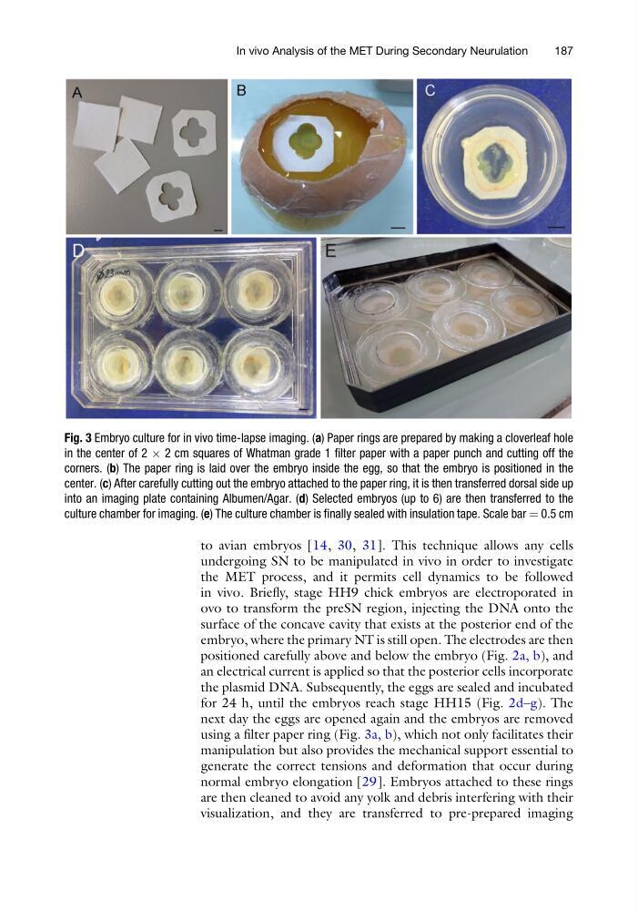
using a filter paper ring (Fig. 3a, b), which not only facilitates their
manipulation but also provides the mechanical support essential to
generate the correct tensions and deformation that occur during
normal embryo elongation [29]. Embryos attached to these rings
are then cleaned to avoid any yolk and debris interfering with their
visualization, and they are transferred to pre-prepared imaging
Fig. 3 Embryo culture for in vivo time-lapse imaging. (a) Paper rings are prepared by making a cloverleaf hole
in the center of 2 Â 2 cm squares of Whatman grade 1 filter paper with a paper punch and cutting off the
corners. (b) The paper ring is laid over the embryo inside the egg, so that the embryo is positioned in the
center. (c) After carefully cutting out the embryo attached to the paper ring, it is then transferred dorsal side up
into an imaging plate containing Albumen/Agar. (d) Selected embryos (up to 6) are then transferred to the
culture chamber for imaging. (e) The culture chamber is finally sealed with insulation tape. Scale bar ¼ 0.5 cm
In vivo Analysis of the MET During Secondary Neurulation
187
undergoing SN to be manipulated in vivo in order to investigate
the MET process, and it permits cell dynamics to be followed
in vivo. Briefly, stage HH9 chick embryos are electroporated in
ovo to transform the preSN region, injecting the DNA onto the
surface of the concave cavity that exists at the posterior end of the
embryo, where the primary NT is still open. The electrodes are then
positioned carefully above and below the embryo (Fig. 2a, b), and
an electrical current is applied so that the posterior cells incorporate
the plasmid DNA. Subsequently, the eggs are sealed and incubated
for 24 h, until the embryos reach stage HH15 (Fig. 2d–g). The
next day the eggs are opened again and the embryos are removed
using a filter paper ring (Fig. 3a, b), which not only facilitates their
manipulation but also provides the mechanical support essential to
generate the correct tensions and deformation that occur during
normal embryo elongation [29]. Embryos attached to these rings
are then cleaned to avoid any yolk and debris interfering with their
visualization, and they are transferred to pre-prepared imaging
Fig. 3 Embryo culture for in vivo time-lapse imaging. (a) Paper rings are prepared by making a cloverleaf hole
in the center of 2 Â 2 cm squares of Whatman grade 1 filter paper with a paper punch and cutting off the
corners. (b) The paper ring is laid over the embryo inside the egg, so that the embryo is positioned in the
center. (c) After carefully cutting out the embryo attached to the paper ring, it is then transferred dorsal side up
into an imaging plate containing Albumen/Agar. (d) Selected embryos (up to 6) are then transferred to the
culture chamber for imaging. (e) The culture chamber is finally sealed with insulation tape. Scale bar ¼ 0.5 cm
In vivo Analysis of the MET During Secondary Neurulation
187