

3. The pierced fly is placed inside the 0.5 ml Eppendorf, and the
tubes are spun at 2348 Â g for 5 min at 4
C. This can be done
either with individual flies or in small batches.
4. Pipette the solution containing the hemolymph and lysis buffer
into a 96-well microplate.
5. Add substrate to the lysate (as directed by manufacturer of
Luciferase reporter kit).
6. Within 5 min (see Note 6) read the bioluminescence levels on a
microplate reader.
7. Raw luciferase data is then compared with the control conditions. Controls should be read in parallel in each individual
experiment.
4 Notes
1. The guts of males and females as well as virgin females and
mated females are physiologically and morphologically distinct
[7], therefore to keep data constant we normally analyze mated
females. For this reason, a small number of males are added to
the vials, but only the females are analyzed.
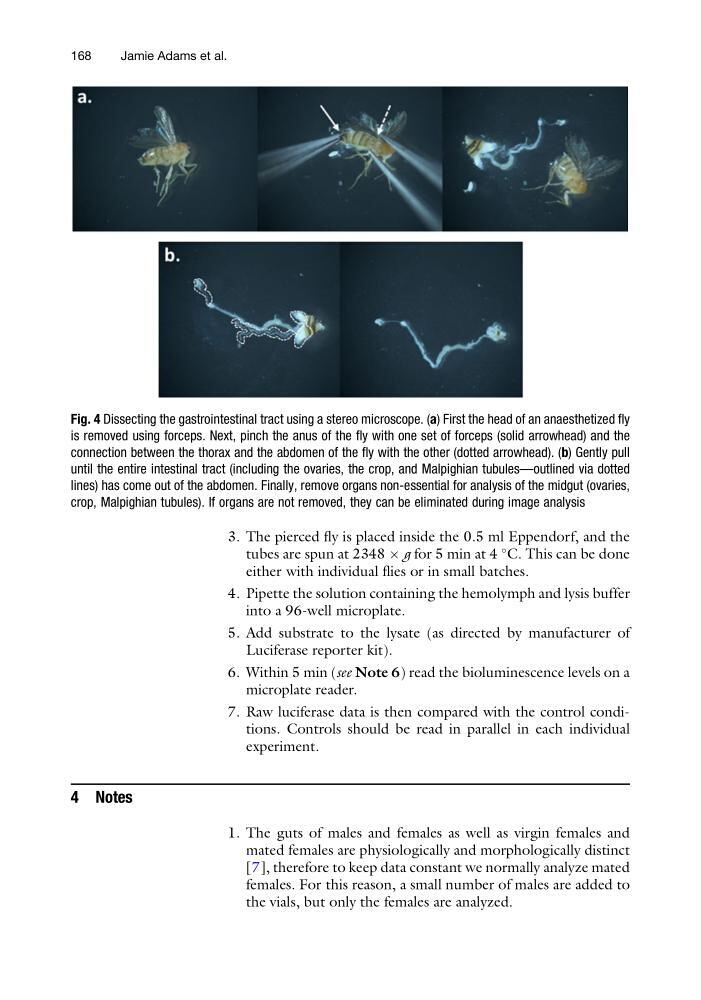
Fig. 4 Dissecting the gastrointestinal tract using a stereo microscope. (a) First the head of an anaesthetized fly
is removed using forceps. Next, pinch the anus of the fly with one set of forceps (solid arrowhead) and the
connection between the thorax and the abdomen of the fly with the other (dotted arrowhead). (b) Gently pull
until the entire intestinal tract (including the ovaries, the crop, and Malpighian tubules—outlined via dotted
lines) has come out of the abdomen. Finally, remove organs non-essential for analysis of the midgut (ovaries,
crop, Malpighian tubules). If organs are not removed, they can be eliminated during image analysis
168
Jamie Adams et al.
tubes are spun at 2348 Â g for 5 min at 4
C. This can be done
either with individual flies or in small batches.
4. Pipette the solution containing the hemolymph and lysis buffer
into a 96-well microplate.
5. Add substrate to the lysate (as directed by manufacturer of
Luciferase reporter kit).
6. Within 5 min (see Note 6) read the bioluminescence levels on a
microplate reader.
7. Raw luciferase data is then compared with the control conditions. Controls should be read in parallel in each individual
experiment.
4 Notes
1. The guts of males and females as well as virgin females and
mated females are physiologically and morphologically distinct
[7], therefore to keep data constant we normally analyze mated
females. For this reason, a small number of males are added to
the vials, but only the females are analyzed.
Fig. 4 Dissecting the gastrointestinal tract using a stereo microscope. (a) First the head of an anaesthetized fly
is removed using forceps. Next, pinch the anus of the fly with one set of forceps (solid arrowhead) and the
connection between the thorax and the abdomen of the fly with the other (dotted arrowhead). (b) Gently pull
until the entire intestinal tract (including the ovaries, the crop, and Malpighian tubules—outlined via dotted
lines) has come out of the abdomen. Finally, remove organs non-essential for analysis of the midgut (ovaries,
crop, Malpighian tubules). If organs are not removed, they can be eliminated during image analysis
168
Jamie Adams et al.