

3. Remove a fraction of the liquid for analysis and pipette into a
96-well microplate.
4. Add substrate to the lysate (as directed by manufacturer of
Luciferase reporter kit).
5. Within 5 min (see Note 6) read the bioluminescence levels on a
microplate reader.
6. Raw luciferase data is then compared with the control conditions. Controls should be read in parallel in each individual
experiment.
3.5 High-Throughput
Analysis of Circulating
Tumor Cell Number
1. Hemolymph is extracted from whole adult flies according to
the instructional video published by Laura Musselman
(https://www.youtube.com/watch?v¼im78OIBKlPA). The
fly abdomen is pierced where it meets the thorax with a tungsten needle.
2. A 0.5 ml Eppendorf is pierced with an x needle, the lid is cut off
and placed in a 1.5 ml Eppendorf containing lysis buffer.
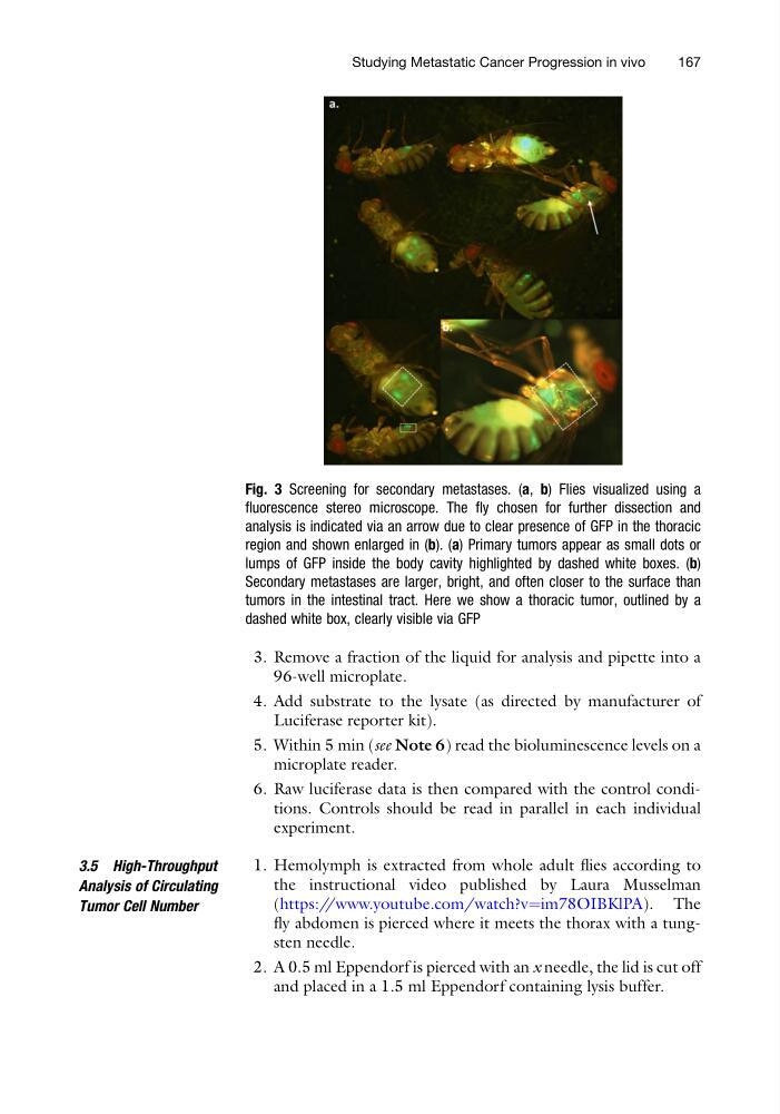
Fig. 3 Screening for secondary metastases. (a, b) Flies visualized using a
fluorescence stereo microscope. The fly chosen for further dissection and
analysis is indicated via an arrow due to clear presence of GFP in the thoracic
region and shown enlarged in (b). (a) Primary tumors appear as small dots or
lumps of GFP inside the body cavity highlighted by dashed white boxes. (b)
Secondary metastases are larger, bright, and often closer to the surface than
tumors in the intestinal tract. Here we show a thoracic tumor, outlined by a
dashed white box, clearly visible via GFP
Studying Metastatic Cancer Progression in vivo
167
96-well microplate.
4. Add substrate to the lysate (as directed by manufacturer of
Luciferase reporter kit).
5. Within 5 min (see Note 6) read the bioluminescence levels on a
microplate reader.
6. Raw luciferase data is then compared with the control conditions. Controls should be read in parallel in each individual
experiment.
3.5 High-Throughput
Analysis of Circulating
Tumor Cell Number
1. Hemolymph is extracted from whole adult flies according to
the instructional video published by Laura Musselman
(https://www.youtube.com/watch?v¼im78OIBKlPA). The
fly abdomen is pierced where it meets the thorax with a tungsten needle.
2. A 0.5 ml Eppendorf is pierced with an x needle, the lid is cut off
and placed in a 1.5 ml Eppendorf containing lysis buffer.
Fig. 3 Screening for secondary metastases. (a, b) Flies visualized using a
fluorescence stereo microscope. The fly chosen for further dissection and
analysis is indicated via an arrow due to clear presence of GFP in the thoracic
region and shown enlarged in (b). (a) Primary tumors appear as small dots or
lumps of GFP inside the body cavity highlighted by dashed white boxes. (b)
Secondary metastases are larger, bright, and often closer to the surface than
tumors in the intestinal tract. Here we show a thoracic tumor, outlined by a
dashed white box, clearly visible via GFP
Studying Metastatic Cancer Progression in vivo
167