

2. The MpTs become very fragile when they have tumors, and
prone to breaking. They should be dissected with care and
checked to make sure they are intact. This is to ensure that
MpTs tumors are not mistaken for secondary tumors.
3. To fix adult midguts (or secondary metastases), incubate in
PBS and 4% electron microscope grade formaldehyde for
30 min. Rinse the samples three times with PBT-BSA (PBS,
4% BSA, 0.1% Triton X-100). Next, incubate with the primary
antibody overnight at 4
C followed by agitation with the
secondary antibody for 2 h at room temperature. Finally,
rinse the samples three times with PBT-BSA and mount in an
appropriate mounting medium such as Vectashield or
Fluoromount.
4. The ureter and base of the MpTs are prone to large bloated
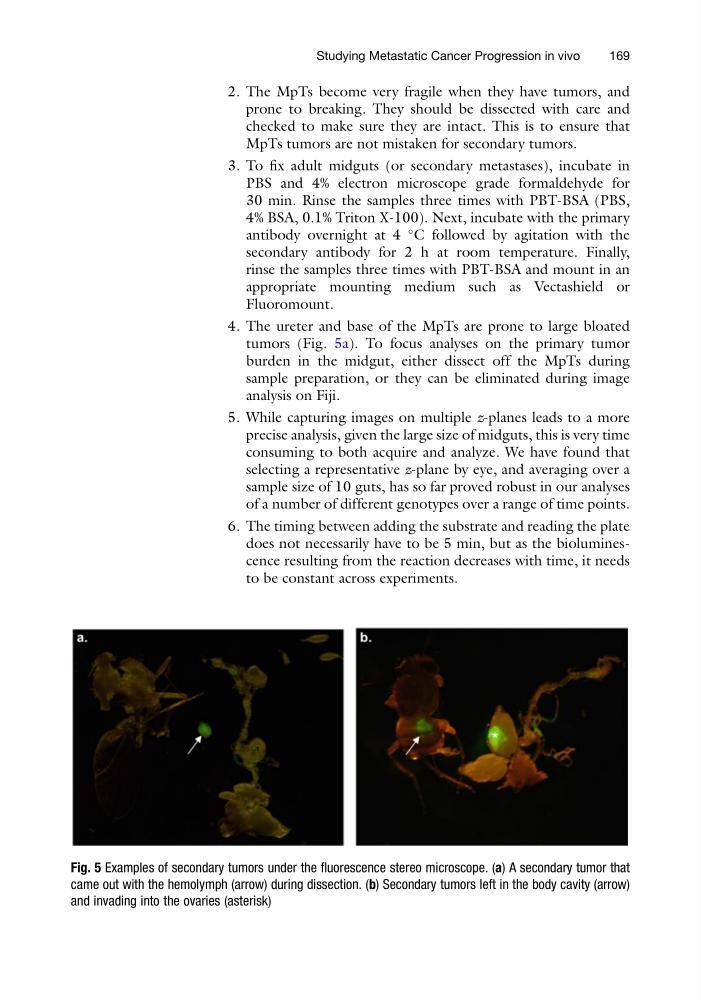
tumors (Fig. 5a). To focus analyses on the primary tumor
burden in the midgut, either dissect off the MpTs during
sample preparation, or they can be eliminated during image
analysis on Fiji.
5. While capturing images on multiple z-planes leads to a more
precise analysis, given the large size of midguts, this is very time
consuming to both acquire and analyze. We have found that
selecting a representative z-plane by eye, and averaging over a
sample size of 10 guts, has so far proved robust in our analyses
of a number of different genotypes over a range of time points.
6. The timing between adding the substrate and reading the plate
does not necessarily have to be 5 min, but as the bioluminescence resulting from the reaction decreases with time, it needs
to be constant across experiments.
Fig. 5 Examples of secondary tumors under the fluorescence stereo microscope. (a) A secondary tumor that
came out with the hemolymph (arrow) during dissection. (b) Secondary tumors left in the body cavity (arrow)
and invading into the ovaries (asterisk)
Studying Metastatic Cancer Progression in vivo
169
prone to breaking. They should be dissected with care and
checked to make sure they are intact. This is to ensure that
MpTs tumors are not mistaken for secondary tumors.
3. To fix adult midguts (or secondary metastases), incubate in
PBS and 4% electron microscope grade formaldehyde for
30 min. Rinse the samples three times with PBT-BSA (PBS,
4% BSA, 0.1% Triton X-100). Next, incubate with the primary
antibody overnight at 4
C followed by agitation with the
secondary antibody for 2 h at room temperature. Finally,
rinse the samples three times with PBT-BSA and mount in an
appropriate mounting medium such as Vectashield or
Fluoromount.
4. The ureter and base of the MpTs are prone to large bloated
tumors (Fig. 5a). To focus analyses on the primary tumor
burden in the midgut, either dissect off the MpTs during
sample preparation, or they can be eliminated during image
analysis on Fiji.
5. While capturing images on multiple z-planes leads to a more
precise analysis, given the large size of midguts, this is very time
consuming to both acquire and analyze. We have found that
selecting a representative z-plane by eye, and averaging over a
sample size of 10 guts, has so far proved robust in our analyses
of a number of different genotypes over a range of time points.
6. The timing between adding the substrate and reading the plate
does not necessarily have to be 5 min, but as the bioluminescence resulting from the reaction decreases with time, it needs
to be constant across experiments.
Fig. 5 Examples of secondary tumors under the fluorescence stereo microscope. (a) A secondary tumor that
came out with the hemolymph (arrow) during dissection. (b) Secondary tumors left in the body cavity (arrow)
and invading into the ovaries (asterisk)
Studying Metastatic Cancer Progression in vivo
169