

4. Embryos should be staged according to anatomical landmarks
[15], as there is great shape and size variability between and
even within litters dissected at a defined time.
5. Speed is essential during dissection, so a learning curve is to be
expected before embryo survival is optimal. It is best to dissect
two or three deciduae, transfer embryos in the incubator, and
then proceed to the next batch.
6. Options include reducing laser power (use minimum laser
power to be able to distinguish cell shape, not to obtain a
perfect image. Signal-noise ratio can be optimized though
post-imaging processing); reducing the time embryos are
exposed to the laser (by decreasing the frequency of scans,
increasing the size of optical sections and/or scan speed). For
the latter, imaging two fluorescent labels is done on one channel track, 1024 Â 1024 resolution at maximal scanning speed
and 2Â signal averaging. Since only 1/3 to 1/4 of the cells are
labeled, cells can be easily tracked even when imaged every
20 min. Cell shape changes are progressive, so we rarely miss
essential events at this rate. 3 μm intervals between individual
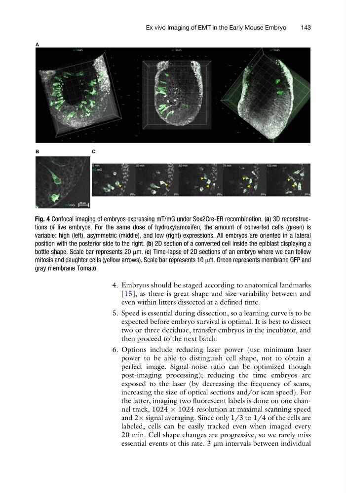
Fig. 4 Confocal imaging of embryos expressing mT/mG under Sox2Cre-ER recombination. (a) 3D reconstructions of live embryos. For the same dose of hydroxytamoxifen, the amount of converted cells (green) is
variable: high (left), asymmetric (middle), and low (right) expressions. All embryos are oriented in a lateral
position with the posterior side to the right. (b) 2D section of a converted cell inside the epiblast displaying a
bottle shape. Scale bar represents 20 μm. (c) Time-lapse of 2D sections of an embryo where we can follow
mitosis and daughter cells (yellow arrows). Scale bar represents 10 μm. Green represents membrane GFP and
gray membrane Tomato
Ex vivo Imaging of EMT in the Early Mouse Embryo
143
[15], as there is great shape and size variability between and
even within litters dissected at a defined time.
5. Speed is essential during dissection, so a learning curve is to be
expected before embryo survival is optimal. It is best to dissect
two or three deciduae, transfer embryos in the incubator, and
then proceed to the next batch.
6. Options include reducing laser power (use minimum laser
power to be able to distinguish cell shape, not to obtain a
perfect image. Signal-noise ratio can be optimized though
post-imaging processing); reducing the time embryos are
exposed to the laser (by decreasing the frequency of scans,
increasing the size of optical sections and/or scan speed). For
the latter, imaging two fluorescent labels is done on one channel track, 1024 Â 1024 resolution at maximal scanning speed
and 2Â signal averaging. Since only 1/3 to 1/4 of the cells are
labeled, cells can be easily tracked even when imaged every
20 min. Cell shape changes are progressive, so we rarely miss
essential events at this rate. 3 μm intervals between individual
Fig. 4 Confocal imaging of embryos expressing mT/mG under Sox2Cre-ER recombination. (a) 3D reconstructions of live embryos. For the same dose of hydroxytamoxifen, the amount of converted cells (green) is
variable: high (left), asymmetric (middle), and low (right) expressions. All embryos are oriented in a lateral
position with the posterior side to the right. (b) 2D section of a converted cell inside the epiblast displaying a
bottle shape. Scale bar represents 20 μm. (c) Time-lapse of 2D sections of an embryo where we can follow
mitosis and daughter cells (yellow arrows). Scale bar represents 10 μm. Green represents membrane GFP and
gray membrane Tomato
Ex vivo Imaging of EMT in the Early Mouse Embryo
143