

4 Notes
1. It is essential to culture embryos in a CO 2 controlled environment (5%) as HEPES-buffered media are toxic for the embryos
when used for long periods of time. It is ideal to culture E6.5–
E8 embryos at 5% O 2 [8] . However, embryos grow adequately
in 5% CO 2 in atmosphere (20% O 2 ) for the usual imaging time
frame (8–12 h) [7].
2. Mouse lines are backcrossed into an outbred CD1 background
as it results in bigger litter sizes, and bigger embryos for a
similar morphological stage that display better resistance to
phototoxicity.
3. Introduction of tamoxifen is toxic even at low concentration
before E4.5, and embryonic toxicity is dose-dependent
between E5.5 and E7.5. In our system, hydroxytamoxifen
quantities (0.1 mg/g) sufficient to trigger efficient recombination could only be injected from E6.25 onwards while preserving embryo health.
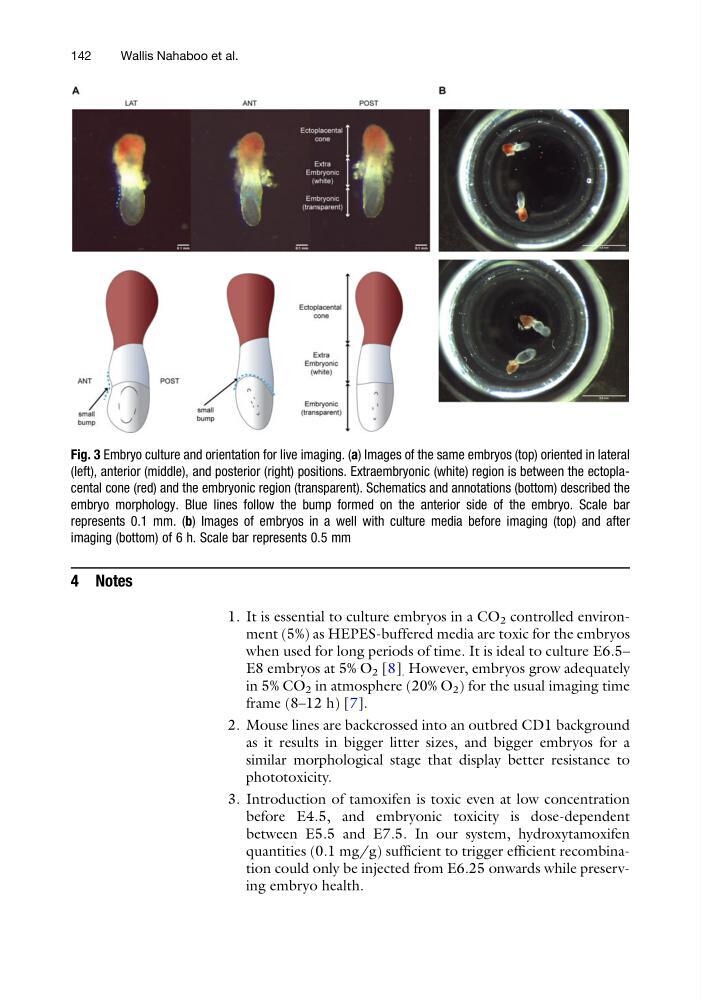
Fig. 3 Embryo culture and orientation for live imaging. (a) Images of the same embryos (top) oriented in lateral
(left), anterior (middle), and posterior (right) positions. Extraembryonic (white) region is between the ectoplacental cone (red) and the embryonic region (transparent). Schematics and annotations (bottom) described the
embryo morphology. Blue lines follow the bump formed on the anterior side of the embryo. Scale bar
represents 0.1 mm. (b) Images of embryos in a well with culture media before imaging (top) and after
imaging (bottom) of 6 h. Scale bar represents 0.5 mm
142
Wallis Nahaboo et al.
1. It is essential to culture embryos in a CO 2 controlled environment (5%) as HEPES-buffered media are toxic for the embryos
when used for long periods of time. It is ideal to culture E6.5–
E8 embryos at 5% O 2 [8] . However, embryos grow adequately
in 5% CO 2 in atmosphere (20% O 2 ) for the usual imaging time
frame (8–12 h) [7].
2. Mouse lines are backcrossed into an outbred CD1 background
as it results in bigger litter sizes, and bigger embryos for a
similar morphological stage that display better resistance to
phototoxicity.
3. Introduction of tamoxifen is toxic even at low concentration
before E4.5, and embryonic toxicity is dose-dependent
between E5.5 and E7.5. In our system, hydroxytamoxifen
quantities (0.1 mg/g) sufficient to trigger efficient recombination could only be injected from E6.25 onwards while preserving embryo health.
Fig. 3 Embryo culture and orientation for live imaging. (a) Images of the same embryos (top) oriented in lateral
(left), anterior (middle), and posterior (right) positions. Extraembryonic (white) region is between the ectoplacental cone (red) and the embryonic region (transparent). Schematics and annotations (bottom) described the
embryo morphology. Blue lines follow the bump formed on the anterior side of the embryo. Scale bar
represents 0.1 mm. (b) Images of embryos in a well with culture media before imaging (top) and after
imaging (bottom) of 6 h. Scale bar represents 0.5 mm
142
Wallis Nahaboo et al.