

186
7 Molecular Structures from Gas-Phase Electron Diffraction
Table 7.2 Total vibrational corrections r tot to the experimental internuclear distances r g and
centrifugal distortion corrections δ cent for 2-cyanopyridine (all values in pm) (Vogt et al. 2019)
Bonds a
r se
e
b,c
δ cent
r 1 d
r 2 d
r 3 d
r tot = r g − r se
e
C7≡N8
115.8(5)
0.0 0
1.8 6
−1.8 1
0.5 7
0.6 2
N1–C6
133.2(2) 1
0.0 0
0.5 7
−0.4 8
0.6 8
0.7 7
N1–C2
133.7(2) 1
0.0 0
0.3 4
−0.2 4
0.6 5
0.7 5
C4–C5
138.4(2) 2
0.0 0
0.4 6
−0.3 7
0.7 5
0.8 4
C3–C4
138.7(2) 2
0.0 0
0.6 4
−0.5 3
0.7 5
0.8 6
C5–C6
139.1(2) 2
0.0 0
0.4 8
−0.3 9
0.7 6
0.8 5
C2–C3
139.3(2) 2
0.0 2
0.3 4
−0.2 4
0.7 4
0.8 5
C2–C7
144.5(3)
0.0 0
0.4 5
−0.3 1
0.6 9
0.8 3
C3–H
107.9(6) 3
0.0 0
1.8 3
−1.4 5
1.6 9
2.0 7
C5–H
108.0(6) 3
0.0 0
2.0 7
−1.6 9
1.6 9
2.0 7
C4–H
108.0(6) 3
0.0 0
1.8 4
−1.4 5
1.6 9
2.0 8
C6–H
108.1(6) 3
0.0 1
1.7 3
−1.3 3
1.7 1
2.1 2
Reprinted by permission from Springer Nature: Springer, Structural Chemistry. The equilibrium
molecular structure of 2-cyanopyridine from combined analysis of gas-phase electron diffraction
and microwave data and results of ab initio calculations; Vogt N, Khaikin LS, Rykov AN, Grikina
OE, Batiukov AA, Vogt J, Kochikov IV, Shishkov IF, 30:1699–1706, copyright 2019
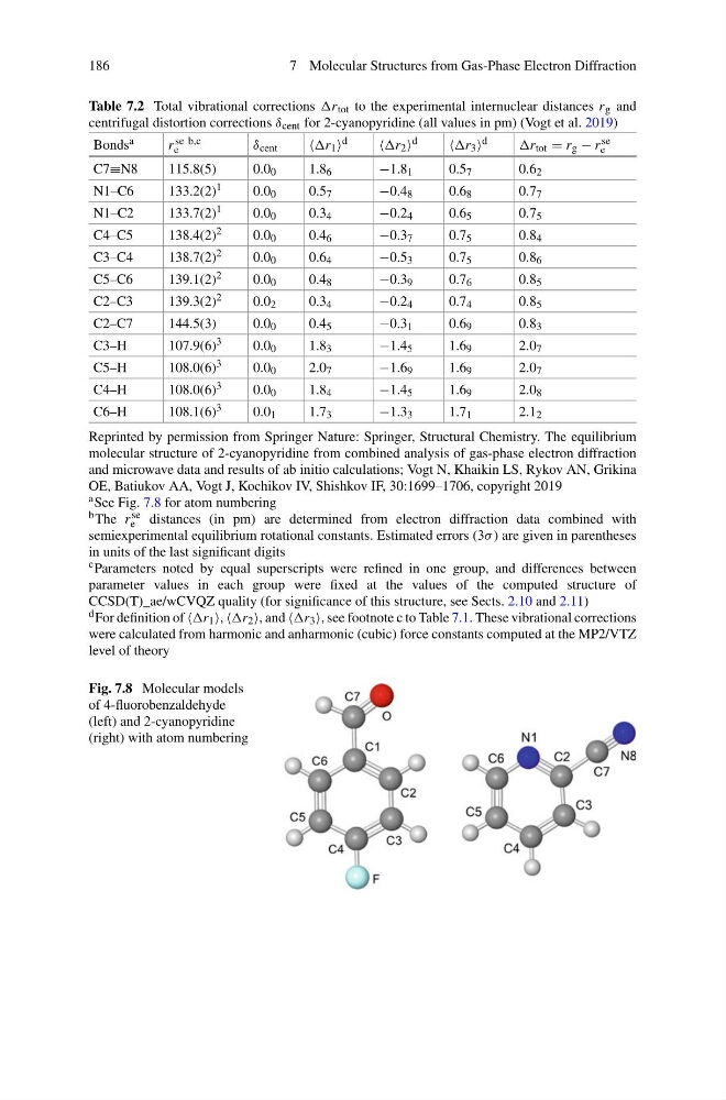
a See Fig. 7.8 for atom numbering
b The r se
e distances (in pm) are determined from electron diffraction data combined with
semiexperimental equilibrium rotational constants. Estimated errors (3σ ) are given in parentheses
in units of the last significant digits
c Parameters noted by equal superscripts were refined in one group, and differences between
parameter values in each group were fixed at the values of the computed structure of
CCSD(T)_ae/wCVQZ quality (for significance of this structure, see Sects. 2.10 and 2.11)
d For definition of r 1 , r 2 , and r 3 , see footnote c to Table 7.1. These vibrational corrections
were calculated from harmonic and anharmonic (cubic) force constants computed at the MP2/VTZ
level of theory
Fig. 7.8 Molecular models
of 4-fluorobenzaldehyde
(left) and 2-cyanopyridine
(right) with atom numbering
7 Molecular Structures from Gas-Phase Electron Diffraction
Table 7.2 Total vibrational corrections r tot to the experimental internuclear distances r g and
centrifugal distortion corrections δ cent for 2-cyanopyridine (all values in pm) (Vogt et al. 2019)
Bonds a
r se
e
b,c
δ cent
r 1 d
r 2 d
r 3 d
r tot = r g − r se
e
C7≡N8
115.8(5)
0.0 0
1.8 6
−1.8 1
0.5 7
0.6 2
N1–C6
133.2(2) 1
0.0 0
0.5 7
−0.4 8
0.6 8
0.7 7
N1–C2
133.7(2) 1
0.0 0
0.3 4
−0.2 4
0.6 5
0.7 5
C4–C5
138.4(2) 2
0.0 0
0.4 6
−0.3 7
0.7 5
0.8 4
C3–C4
138.7(2) 2
0.0 0
0.6 4
−0.5 3
0.7 5
0.8 6
C5–C6
139.1(2) 2
0.0 0
0.4 8
−0.3 9
0.7 6
0.8 5
C2–C3
139.3(2) 2
0.0 2
0.3 4
−0.2 4
0.7 4
0.8 5
C2–C7
144.5(3)
0.0 0
0.4 5
−0.3 1
0.6 9
0.8 3
C3–H
107.9(6) 3
0.0 0
1.8 3
−1.4 5
1.6 9
2.0 7
C5–H
108.0(6) 3
0.0 0
2.0 7
−1.6 9
1.6 9
2.0 7
C4–H
108.0(6) 3
0.0 0
1.8 4
−1.4 5
1.6 9
2.0 8
C6–H
108.1(6) 3
0.0 1
1.7 3
−1.3 3
1.7 1
2.1 2
Reprinted by permission from Springer Nature: Springer, Structural Chemistry. The equilibrium
molecular structure of 2-cyanopyridine from combined analysis of gas-phase electron diffraction
and microwave data and results of ab initio calculations; Vogt N, Khaikin LS, Rykov AN, Grikina
OE, Batiukov AA, Vogt J, Kochikov IV, Shishkov IF, 30:1699–1706, copyright 2019
a See Fig. 7.8 for atom numbering
b The r se
e distances (in pm) are determined from electron diffraction data combined with
semiexperimental equilibrium rotational constants. Estimated errors (3σ ) are given in parentheses
in units of the last significant digits
c Parameters noted by equal superscripts were refined in one group, and differences between
parameter values in each group were fixed at the values of the computed structure of
CCSD(T)_ae/wCVQZ quality (for significance of this structure, see Sects. 2.10 and 2.11)
d For definition of r 1 , r 2 , and r 3 , see footnote c to Table 7.1. These vibrational corrections
were calculated from harmonic and anharmonic (cubic) force constants computed at the MP2/VTZ
level of theory
Fig. 7.8 Molecular models
of 4-fluorobenzaldehyde
(left) and 2-cyanopyridine
(right) with atom numbering