

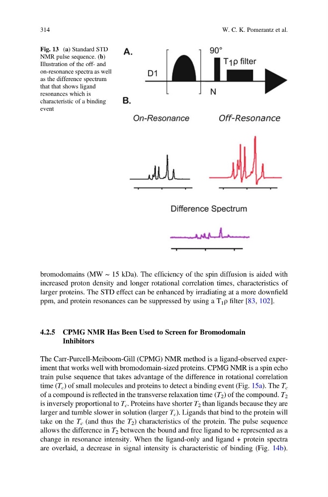
bromodomains (MW ~ 15 kDa). The efficiency of the spin diffusion is aided with
increased proton density and longer rotational correlation times, characteristics of
larger proteins. The STD effect can be enhanced by irradiating at a more downfield
ppm, and protein resonances can be suppressed by using a T 1 ρ filter [83, 102].
4.2.5 CPMG NMR Has Been Used to Screen for Bromodomain
Inhibitors
The Carr-Purcell-Meiboom-Gill (CPMG) NMR method is a ligand-observed experiment that works well with bromodomain-sized proteins. CPMG NMR is a spin echo
train pulse sequence that takes advantage of the difference in rotational correlation
time (T c ) of small molecules and proteins to detect a binding event (Fig. 15a). The T c
of a compound is reflected in the transverse relaxation time (T 2 ) of the compound. T 2
is inversely proportional to T c . Proteins have shorter T 2 than ligands because they are
larger and tumble slower in solution (larger T c ). Ligands that bind to the protein will
take on the T c (and thus the T 2 ) characteristics of the protein. The pulse sequence
allows the difference in T 2 between the bound and free ligand to be represented as a
change in resonance intensity. When the ligand-only and ligand + protein spectra
are overlaid, a decrease in signal intensity is characteristic of binding (Fig. 14b).
Fig. 13 (a) Standard STD
NMR pulse sequence. (b)
Illustration of the off- and
on-resonance spectra as well
as the difference spectrum
that that shows ligand
resonances which is
characteristic of a binding
event
314
W. C. K. Pomerantz et al.
increased proton density and longer rotational correlation times, characteristics of
larger proteins. The STD effect can be enhanced by irradiating at a more downfield
ppm, and protein resonances can be suppressed by using a T 1 ρ filter [83, 102].
4.2.5 CPMG NMR Has Been Used to Screen for Bromodomain
Inhibitors
The Carr-Purcell-Meiboom-Gill (CPMG) NMR method is a ligand-observed experiment that works well with bromodomain-sized proteins. CPMG NMR is a spin echo
train pulse sequence that takes advantage of the difference in rotational correlation
time (T c ) of small molecules and proteins to detect a binding event (Fig. 15a). The T c
of a compound is reflected in the transverse relaxation time (T 2 ) of the compound. T 2
is inversely proportional to T c . Proteins have shorter T 2 than ligands because they are
larger and tumble slower in solution (larger T c ). Ligands that bind to the protein will
take on the T c (and thus the T 2 ) characteristics of the protein. The pulse sequence
allows the difference in T 2 between the bound and free ligand to be represented as a
change in resonance intensity. When the ligand-only and ligand + protein spectra
are overlaid, a decrease in signal intensity is characteristic of binding (Fig. 14b).
Fig. 13 (a) Standard STD
NMR pulse sequence. (b)
Illustration of the off- and
on-resonance spectra as well
as the difference spectrum
that that shows ligand
resonances which is
characteristic of a binding
event
314
W. C. K. Pomerantz et al.