

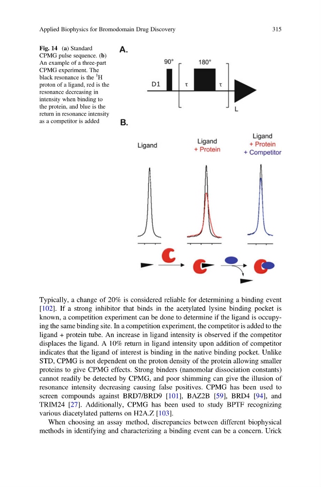
Typically, a change of 20% is considered reliable for determining a binding event
[102]. If a strong inhibitor that binds in the acetylated lysine binding pocket is
known, a competition experiment can be done to determine if the ligand is occupying the same binding site. In a competition experiment, the competitor is added to the
ligand + protein tube. An increase in ligand intensity is observed if the competitor
displaces the ligand. A 10% return in ligand intensity upon addition of competitor
indicates that the ligand of interest is binding in the native binding pocket. Unlike
STD, CPMG is not dependent on the proton density of the protein allowing smaller
proteins to give CPMG effects. Strong binders (nanomolar dissociation constants)
cannot readily be detected by CPMG, and poor shimming can give the illusion of
resonance intensity decreasing causing false positives. CPMG has been used to
screen compounds against BRD7/BRD9 [101], BAZ2B [59], BRD4 [94], and
TRIM24 [27]. Additionally, CPMG has been used to study BPTF recognizing
various diacetylated patterns on H2A.Z [103].
When choosing an assay method, discrepancies between different biophysical
methods in identifying and characterizing a binding event can be a concern. Urick
Fig. 14 (a) Standard
CPMG pulse sequence. (b)
An example of a three-part
CPMG experiment. The
black resonance is the
1
H
proton of a ligand, red is the
resonance decreasing in
intensity when binding to
the protein, and blue is the
return in resonance intensity
as a competitor is added
Applied Biophysics for Bromodomain Drug Discovery
315
[102]. If a strong inhibitor that binds in the acetylated lysine binding pocket is
known, a competition experiment can be done to determine if the ligand is occupying the same binding site. In a competition experiment, the competitor is added to the
ligand + protein tube. An increase in ligand intensity is observed if the competitor
displaces the ligand. A 10% return in ligand intensity upon addition of competitor
indicates that the ligand of interest is binding in the native binding pocket. Unlike
STD, CPMG is not dependent on the proton density of the protein allowing smaller
proteins to give CPMG effects. Strong binders (nanomolar dissociation constants)
cannot readily be detected by CPMG, and poor shimming can give the illusion of
resonance intensity decreasing causing false positives. CPMG has been used to
screen compounds against BRD7/BRD9 [101], BAZ2B [59], BRD4 [94], and
TRIM24 [27]. Additionally, CPMG has been used to study BPTF recognizing
various diacetylated patterns on H2A.Z [103].
When choosing an assay method, discrepancies between different biophysical
methods in identifying and characterizing a binding event can be a concern. Urick
Fig. 14 (a) Standard
CPMG pulse sequence. (b)
An example of a three-part
CPMG experiment. The
black resonance is the
1
H
proton of a ligand, red is the
resonance decreasing in
intensity when binding to
the protein, and blue is the
return in resonance intensity
as a competitor is added
Applied Biophysics for Bromodomain Drug Discovery
315