

TET1 and TET3 have an additional DNA-binding CXXC domain at the N-terminus,
which preferentially binds CpG-rich regions. TET2 lacks the CXXC zinc finger
domain but instead has been shown to interact with IDAX (CXXC4) which then
interacts with DNA [6, 114].
While the cysteine-rich domain is essential for the catalytic activity [114],
truncations in LCI retain the activity in all hTET1–3, albeit at reduced catalytic
efficiency [108, 114]. The first structural insight came in 2013 from the crystal
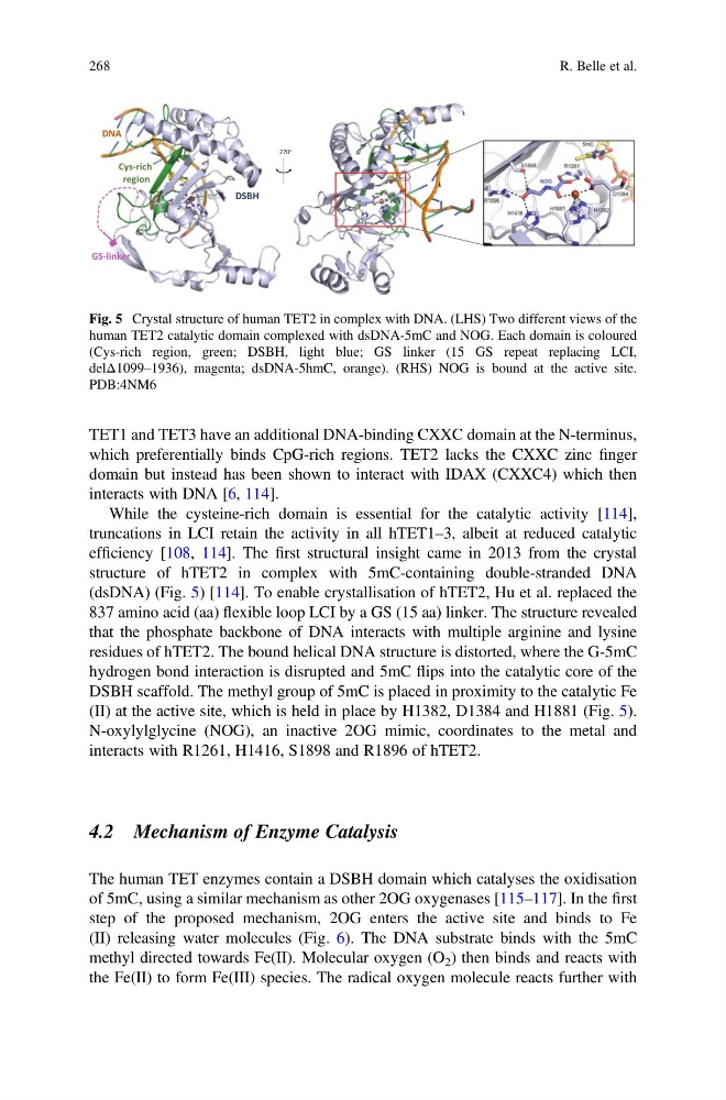
structure of hTET2 in complex with 5mC-containing double-stranded DNA
(dsDNA) (Fig. 5) [114]. To enable crystallisation of hTET2, Hu et al. replaced the
837 amino acid (aa) flexible loop LCI by a GS (15 aa) linker. The structure revealed
that the phosphate backbone of DNA interacts with multiple arginine and lysine
residues of hTET2. The bound helical DNA structure is distorted, where the G-5mC
hydrogen bond interaction is disrupted and 5mC flips into the catalytic core of the
DSBH scaffold. The methyl group of 5mC is placed in proximity to the catalytic Fe
(II) at the active site, which is held in place by H1382, D1384 and H1881 (Fig. 5).
N-oxylylglycine (NOG), an inactive 2OG mimic, coordinates to the metal and
interacts with R1261, H1416, S1898 and R1896 of hTET2.
4.2 Mechanism of Enzyme Catalysis
The human TET enzymes contain a DSBH domain which catalyses the oxidisation
of 5mC, using a similar mechanism as other 2OG oxygenases [115–117]. In the first
step of the proposed mechanism, 2OG enters the active site and binds to Fe
(II) releasing water molecules (Fig. 6). The DNA substrate binds with the 5mC
methyl directed towards Fe(II). Molecular oxygen (O 2 ) then binds and reacts with
the Fe(II) to form Fe(III) species. The radical oxygen molecule reacts further with
270 ∘
Cys-rich
region
DSBH
DNA
GS-linker
Fig. 5 Crystal structure of human TET2 in complex with DNA. (LHS) Two different views of the
human TET2 catalytic domain complexed with dsDNA-5mC and NOG. Each domain is coloured
(Cys-rich region, green; DSBH, light blue; GS linker (15 GS repeat replacing LCI,
delΔ1099–1936), magenta; dsDNA-5hmC, orange). (RHS) NOG is bound at the active site.
PDB:4NM6
268
R. Belle et al.
which preferentially binds CpG-rich regions. TET2 lacks the CXXC zinc finger
domain but instead has been shown to interact with IDAX (CXXC4) which then
interacts with DNA [6, 114].
While the cysteine-rich domain is essential for the catalytic activity [114],
truncations in LCI retain the activity in all hTET1–3, albeit at reduced catalytic
efficiency [108, 114]. The first structural insight came in 2013 from the crystal
structure of hTET2 in complex with 5mC-containing double-stranded DNA
(dsDNA) (Fig. 5) [114]. To enable crystallisation of hTET2, Hu et al. replaced the
837 amino acid (aa) flexible loop LCI by a GS (15 aa) linker. The structure revealed
that the phosphate backbone of DNA interacts with multiple arginine and lysine
residues of hTET2. The bound helical DNA structure is distorted, where the G-5mC
hydrogen bond interaction is disrupted and 5mC flips into the catalytic core of the
DSBH scaffold. The methyl group of 5mC is placed in proximity to the catalytic Fe
(II) at the active site, which is held in place by H1382, D1384 and H1881 (Fig. 5).
N-oxylylglycine (NOG), an inactive 2OG mimic, coordinates to the metal and
interacts with R1261, H1416, S1898 and R1896 of hTET2.
4.2 Mechanism of Enzyme Catalysis
The human TET enzymes contain a DSBH domain which catalyses the oxidisation
of 5mC, using a similar mechanism as other 2OG oxygenases [115–117]. In the first
step of the proposed mechanism, 2OG enters the active site and binds to Fe
(II) releasing water molecules (Fig. 6). The DNA substrate binds with the 5mC
methyl directed towards Fe(II). Molecular oxygen (O 2 ) then binds and reacts with
the Fe(II) to form Fe(III) species. The radical oxygen molecule reacts further with
270 ∘
Cys-rich
region
DSBH
DNA
GS-linker
Fig. 5 Crystal structure of human TET2 in complex with DNA. (LHS) Two different views of the
human TET2 catalytic domain complexed with dsDNA-5mC and NOG. Each domain is coloured
(Cys-rich region, green; DSBH, light blue; GS linker (15 GS repeat replacing LCI,
delΔ1099–1936), magenta; dsDNA-5hmC, orange). (RHS) NOG is bound at the active site.
PDB:4NM6
268
R. Belle et al.