

remodelling after mouse fertilisation, indicating the possibility of an additional
mechanism that would remove 5mC [104]. The TET proteins were originally linked
as being fusion partner of mixed lineage leukaemia (MLL) [105] and later identified
as enzymes related to JPB1 that facilitate the oxidation of thymidine (T) in the
nucleus to 5-hydroxyuracil (5hmU) [106]. The first discovery of 5hmC modification
was made in 2009, where 5hmC was detected in the brain tissue at high abundance
[5]. At the same time, it was revealed that TET enzymes are capable of oxidising
5mC to 5hmC [6]. Shortly after, it was discovered that TET enzymes can iteratively
oxidise 5mC to 5caC [107].
TET enzymes are widely conserved through evolution [111], including in
Naegleria gruberi TET (NgTET). Herein, we focus on studies involving human
TETs (hTETs), with some references to other model organisms, such as murine
TETs (mTET) [110] and NgTETs [112].
To date, three TET enzymes have been found in humans (hTET). The hTET
proteins are approximately 180–230 kDa in length, and all human TETs (hTET1–3)
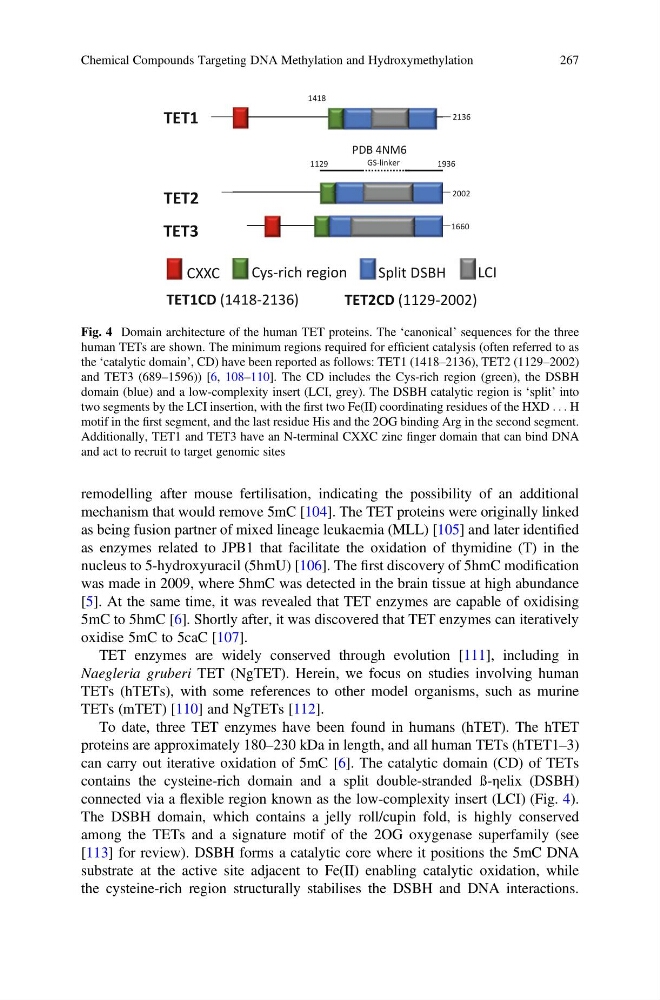
can carry out iterative oxidation of 5mC [6]. The catalytic domain (CD) of TETs
contains the cysteine-rich domain and a split double-stranded ß-ηelix (DSBH)
connected via a flexible region known as the low-complexity insert (LCI) (Fig. 4).
The DSBH domain, which contains a jelly roll/cupin fold, is highly conserved
among the TETs and a signature motif of the 2OG oxygenase superfamily (see
[113] for review). DSBH forms a catalytic core where it positions the 5mC DNA
substrate at the active site adjacent to Fe(II) enabling catalytic oxidation, while
the cysteine-rich region structurally stabilises the DSBH and DNA interactions.
Cys-rich region
CXXC
Split DSBH
LCI
2136
2002
1660
TET1
TET2
TET3
TET1CD (1418-2136)
TET2CD (1129-2002)
Cys-rich region
CXXC
Split DSBH
1418
GS-linker
1129
1936
PDB 4NM6
Fig. 4 Domain architecture of the human TET proteins. The ‘canonical’ sequences for the three
human TETs are shown. The minimum regions required for efficient catalysis (often referred to as
the ‘catalytic domain’, CD) have been reported as follows: TET1 (1418–2136), TET2 (1129–2002)
and TET3 (689–1596)) [6, 108–110]. The CD includes the Cys-rich region (green), the DSBH
domain (blue) and a low-complexity insert (LCI, grey). The DSBH catalytic region is ‘split’ into
two segments by the LCI insertion, with the first two Fe(II) coordinating residues of the HXD . . . H
motif in the first segment, and the last residue His and the 2OG binding Arg in the second segment.
Additionally, TET1 and TET3 have an N-terminal CXXC zinc finger domain that can bind DNA
and act to recruit to target genomic sites
Chemical Compounds Targeting DNA Methylation and Hydroxymethylation
267
mechanism that would remove 5mC [104]. The TET proteins were originally linked
as being fusion partner of mixed lineage leukaemia (MLL) [105] and later identified
as enzymes related to JPB1 that facilitate the oxidation of thymidine (T) in the
nucleus to 5-hydroxyuracil (5hmU) [106]. The first discovery of 5hmC modification
was made in 2009, where 5hmC was detected in the brain tissue at high abundance
[5]. At the same time, it was revealed that TET enzymes are capable of oxidising
5mC to 5hmC [6]. Shortly after, it was discovered that TET enzymes can iteratively
oxidise 5mC to 5caC [107].
TET enzymes are widely conserved through evolution [111], including in
Naegleria gruberi TET (NgTET). Herein, we focus on studies involving human
TETs (hTETs), with some references to other model organisms, such as murine
TETs (mTET) [110] and NgTETs [112].
To date, three TET enzymes have been found in humans (hTET). The hTET
proteins are approximately 180–230 kDa in length, and all human TETs (hTET1–3)
can carry out iterative oxidation of 5mC [6]. The catalytic domain (CD) of TETs
contains the cysteine-rich domain and a split double-stranded ß-ηelix (DSBH)
connected via a flexible region known as the low-complexity insert (LCI) (Fig. 4).
The DSBH domain, which contains a jelly roll/cupin fold, is highly conserved
among the TETs and a signature motif of the 2OG oxygenase superfamily (see
[113] for review). DSBH forms a catalytic core where it positions the 5mC DNA
substrate at the active site adjacent to Fe(II) enabling catalytic oxidation, while
the cysteine-rich region structurally stabilises the DSBH and DNA interactions.
Cys-rich region
CXXC
Split DSBH
LCI
2136
2002
1660
TET1
TET2
TET3
TET1CD (1418-2136)
TET2CD (1129-2002)
Cys-rich region
CXXC
Split DSBH
1418
GS-linker
1129
1936
PDB 4NM6
Fig. 4 Domain architecture of the human TET proteins. The ‘canonical’ sequences for the three
human TETs are shown. The minimum regions required for efficient catalysis (often referred to as
the ‘catalytic domain’, CD) have been reported as follows: TET1 (1418–2136), TET2 (1129–2002)
and TET3 (689–1596)) [6, 108–110]. The CD includes the Cys-rich region (green), the DSBH
domain (blue) and a low-complexity insert (LCI, grey). The DSBH catalytic region is ‘split’ into
two segments by the LCI insertion, with the first two Fe(II) coordinating residues of the HXD . . . H
motif in the first segment, and the last residue His and the 2OG binding Arg in the second segment.
Additionally, TET1 and TET3 have an N-terminal CXXC zinc finger domain that can bind DNA
and act to recruit to target genomic sites
Chemical Compounds Targeting DNA Methylation and Hydroxymethylation
267