

KDM4C [75]. While a co-crystal structure was not obtained, the study highlights
that a small peptide fragment can confer potency and selectivity for JmjCKDMs [75].
3.2.2 Substrate-Binding Pocket Targeted Peptide Inhibitors
mRNA-display-based cyclic peptide library screening against KDM4A generated
potent and selective inhibitors for the KDM4A/B/C family [76]. Interestingly, the
binding cyclic peptides were distinct sequences with no homology to histone H3. A
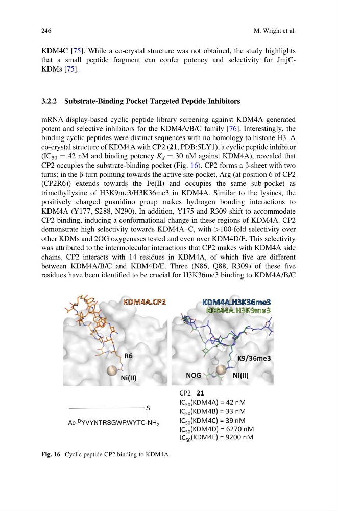
co-crystal structure of KDM4A with CP2 (21, PDB:5LY1), a cyclic peptide inhibitor
(IC 50 ¼ 42 nM and binding potency K d ¼ 30 nM against KDM4A), revealed that
CP2 occupies the substrate-binding pocket (Fig. 16). CP2 forms a β-sheet with two
turns; in the β-turn pointing towards the active site pocket, Arg (at position 6 of CP2
(CP2R6)) extends towards the Fe(II) and occupies the same sub-pocket as
trimethyllysine of H3K9me3/H3K36me3 in KDM4A. Similar to the lysines, the
positively charged guanidino group makes hydrogen bonding interactions to
KDM4A (Y177, S288, N290). In addition, Y175 and R309 shift to accommodate
CP2 binding, inducing a conformational change in these regions of KDM4A. CP2
demonstrate high selectivity towards KDM4A–C, with >100-fold selectivity over
other KDMs and 2OG oxygenases tested and even over KDM4D/E. This selectivity
was attributed to the intermolecular interactions that CP2 makes with KDM4A side
chains. CP2 interacts with 14 residues in KDM4A, of which five are different
between KDM4A/B/C and KDM4D/E. Three (N86, Q88, R309) of these five
residues have been identified to be crucial for H3K36me3 binding to KDM4A/B/C
K9/36me3
R6
Ni(II)
Ni(II)
NOG
CP2 21
IC 50 (KDM4A) = 42 nM
IC 50 (KDM4B) = 33 nM
IC 50 (KDM4C) = 39 nM
(KDM4D) = 6270 nM
IC 50 (KDM4E) = 9200 nM
IC 50
Fig. 16 Cyclic peptide CP2 binding to KDM4A
246
M. Wright et al.
that a small peptide fragment can confer potency and selectivity for JmjCKDMs [75].
3.2.2 Substrate-Binding Pocket Targeted Peptide Inhibitors
mRNA-display-based cyclic peptide library screening against KDM4A generated
potent and selective inhibitors for the KDM4A/B/C family [76]. Interestingly, the
binding cyclic peptides were distinct sequences with no homology to histone H3. A
co-crystal structure of KDM4A with CP2 (21, PDB:5LY1), a cyclic peptide inhibitor
(IC 50 ¼ 42 nM and binding potency K d ¼ 30 nM against KDM4A), revealed that
CP2 occupies the substrate-binding pocket (Fig. 16). CP2 forms a β-sheet with two
turns; in the β-turn pointing towards the active site pocket, Arg (at position 6 of CP2
(CP2R6)) extends towards the Fe(II) and occupies the same sub-pocket as
trimethyllysine of H3K9me3/H3K36me3 in KDM4A. Similar to the lysines, the
positively charged guanidino group makes hydrogen bonding interactions to
KDM4A (Y177, S288, N290). In addition, Y175 and R309 shift to accommodate
CP2 binding, inducing a conformational change in these regions of KDM4A. CP2
demonstrate high selectivity towards KDM4A–C, with >100-fold selectivity over
other KDMs and 2OG oxygenases tested and even over KDM4D/E. This selectivity
was attributed to the intermolecular interactions that CP2 makes with KDM4A side
chains. CP2 interacts with 14 residues in KDM4A, of which five are different
between KDM4A/B/C and KDM4D/E. Three (N86, Q88, R309) of these five
residues have been identified to be crucial for H3K36me3 binding to KDM4A/B/C
K9/36me3
R6
Ni(II)
Ni(II)
NOG
CP2 21
IC 50 (KDM4A) = 42 nM
IC 50 (KDM4B) = 33 nM
IC 50 (KDM4C) = 39 nM
(KDM4D) = 6270 nM
IC 50 (KDM4E) = 9200 nM
IC 50
Fig. 16 Cyclic peptide CP2 binding to KDM4A
246
M. Wright et al.