

[74]. Thus, subtle differences in the substrate-binding pockets can be utilised for the
generation of selective and potent inhibitors against JmjC-KDMs. Structure and
MS-guided modifications of CP2 yielded peptides that retained in vitro potency and
selectivity and increased cellular permeability and cellular KDM4A target engagement, providing promising cellular tools for the KDM4s.
3.2.3 Small Molecule Targeting Substrate-Binding Pocket
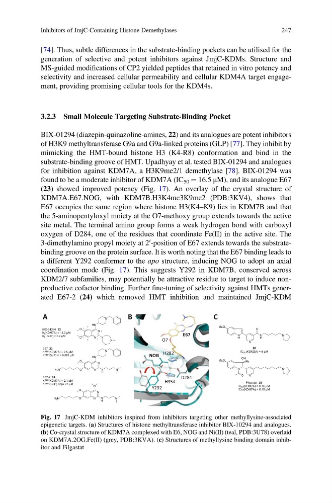
BIX-01294 (diazepin-quinazoline-amines, 22) and its analogues are potent inhibitors
of H3K9 methyltransferase G9a and G9a-linked proteins (GLP) [77]. They inhibit by
mimicking the HMT-bound histone H3 (K4-R8) conformation and bind in the
substrate-binding groove of HMT. Upadhyay et al. tested BIX-01294 and analogues
for inhibition against KDM7A, a H3K9me2/1 demethylase [78]. BIX-01294 was
found to be a moderate inhibitor of KDM7A (IC 50 ¼ 16.5 μM), and its analogue E67
(23) showed improved potency (Fig. 17). An overlay of the crystal structure of
KDM7A.E67.NOG, with KDM7B.H3K4me3K9me2 (PDB:3KV4), shows that
E67 occupies the same region where histone H3(K4–K9) lies in KDM7B and that
the 5-aminopentyloxyl moiety at the O7-methoxy group extends towards the active
site metal. The terminal amino group forms a weak hydrogen bond with carboxyl
oxygen of D284, one of the residues that coordinate Fe(II) in the active site. The
3-dimethylamino propyl moiety at 2
0 -position of E67 extends towards the substratebinding groove on the protein surface. It is worth noting that the E67 binding leads to
a different Y292 conformer to the apo structure, inducing NOG to adopt an axial
coordination mode (Fig. 17). This suggests Y292 in KDM7B, conserved across
KDM2/7 subfamilies, may potentially be attractive residue to target to induce nonproductive cofactor binding. Further fine-tuning of selectivity against HMTs generated E67-2 (24) which removed HMT inhibition and maintained JmjC-KDM
E67
O7
D284
Y292
NOG H282
H354
1
3
5
8
7
6
A
B
C
N
H
MeO
N
O
N
H
N
N
MeO
N
N
O
Ph
CN
Filgastat 25
IC50(KDM2A) = 0.16 μM
IC50(KDM7A) = 0.19 μM
26
IC50(KDM2A) = 5 μM
N
N
HN
N
O
O
N
N
N
N
HN
N
O
O
N
H
N
N
N
N
O
O
N
H
N
BIX-01294 22
Ki(KDM7A) = 15.3 μM
Ki (GLP) = 0.2 μM
E67 23
Ki app (KDM7A) = 2.5 μM
Ki app (GLP) = < 0.001 μM
E67-2 24
Ki app (KDM7A) = 2.1 μM
Ki app (GLP) appx 75 μM
H2N
H2N
Fig. 17 JmjC-KDM inhibitors inspired from inhibitors targeting other methyllysine-associated
epigenetic targets. (a) Structures of histone methyltransferase inhibitor BIX-10294 and analogues.
(b) Co-crystal structure of KDM7A complexed with E6, NOG and Ni(II) (teal, PDB:3U78) overlaid
on KDM7A.2OG.Fe(II) (grey, PDB:3KVA). (c) Structures of methyllysine binding domain inhibitor and Filgastat
Inhibitors of JmjC-Containing Histone Demethylases
247
generation of selective and potent inhibitors against JmjC-KDMs. Structure and
MS-guided modifications of CP2 yielded peptides that retained in vitro potency and
selectivity and increased cellular permeability and cellular KDM4A target engagement, providing promising cellular tools for the KDM4s.
3.2.3 Small Molecule Targeting Substrate-Binding Pocket
BIX-01294 (diazepin-quinazoline-amines, 22) and its analogues are potent inhibitors
of H3K9 methyltransferase G9a and G9a-linked proteins (GLP) [77]. They inhibit by
mimicking the HMT-bound histone H3 (K4-R8) conformation and bind in the
substrate-binding groove of HMT. Upadhyay et al. tested BIX-01294 and analogues
for inhibition against KDM7A, a H3K9me2/1 demethylase [78]. BIX-01294 was
found to be a moderate inhibitor of KDM7A (IC 50 ¼ 16.5 μM), and its analogue E67
(23) showed improved potency (Fig. 17). An overlay of the crystal structure of
KDM7A.E67.NOG, with KDM7B.H3K4me3K9me2 (PDB:3KV4), shows that
E67 occupies the same region where histone H3(K4–K9) lies in KDM7B and that
the 5-aminopentyloxyl moiety at the O7-methoxy group extends towards the active
site metal. The terminal amino group forms a weak hydrogen bond with carboxyl
oxygen of D284, one of the residues that coordinate Fe(II) in the active site. The
3-dimethylamino propyl moiety at 2
0 -position of E67 extends towards the substratebinding groove on the protein surface. It is worth noting that the E67 binding leads to
a different Y292 conformer to the apo structure, inducing NOG to adopt an axial
coordination mode (Fig. 17). This suggests Y292 in KDM7B, conserved across
KDM2/7 subfamilies, may potentially be attractive residue to target to induce nonproductive cofactor binding. Further fine-tuning of selectivity against HMTs generated E67-2 (24) which removed HMT inhibition and maintained JmjC-KDM
E67
O7
D284
Y292
NOG H282
H354
1
3
5
8
7
6
A
B
C
N
H
MeO
N
O
N
H
N
N
MeO
N
N
O
Ph
CN
Filgastat 25
IC50(KDM2A) = 0.16 μM
IC50(KDM7A) = 0.19 μM
26
IC50(KDM2A) = 5 μM
N
N
HN
N
O
O
N
N
N
N
HN
N
O
O
N
H
N
N
N
N
O
O
N
H
N
BIX-01294 22
Ki(KDM7A) = 15.3 μM
Ki (GLP) = 0.2 μM
E67 23
Ki app (KDM7A) = 2.5 μM
Ki app (GLP) = < 0.001 μM
E67-2 24
Ki app (KDM7A) = 2.1 μM
Ki app (GLP) appx 75 μM
H2N
H2N
Fig. 17 JmjC-KDM inhibitors inspired from inhibitors targeting other methyllysine-associated
epigenetic targets. (a) Structures of histone methyltransferase inhibitor BIX-10294 and analogues.
(b) Co-crystal structure of KDM7A complexed with E6, NOG and Ni(II) (teal, PDB:3U78) overlaid
on KDM7A.2OG.Fe(II) (grey, PDB:3KVA). (c) Structures of methyllysine binding domain inhibitor and Filgastat
Inhibitors of JmjC-Containing Histone Demethylases
247