

fivefold to tenfold selective over KDM5B and KDM5C [72]. Similar IC 50 s (3–7 μM)
were also obtained with GSK-J4 in cells transfected with KDM6B, KDM4C and
KDM5B. As a result, inhibition by GSK-J1 is likely a combination of activity
towards H3K4me3/me2 and H3K27me3/me2. Despite this, GSK-J1 is the mostselective KDM6B inhibitor and is a useful chemical tool for studying KDM6B
biology.
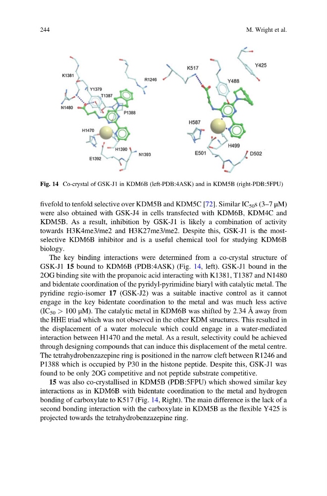
The key binding interactions were determined from a co-crystal structure of
GSK-J1 15 bound to KDM6B (PDB:4ASK) (Fig. 14, left). GSK-J1 bound in the
2OG binding site with the propanoic acid interacting with K1381, T1387 and N1480
and bidentate coordination of the pyridyl-pyrimidine biaryl with catalytic metal. The
pyridine regio-isomer 17 (GSK-J2) was a suitable inactive control as it cannot
engage in the key bidentate coordination to the metal and was much less active
(IC 50 > 100 μM). The catalytic metal in KDM6B was shifted by 2.34 Å away from
the HHE triad which was not observed in the other KDM structures. This resulted in
the displacement of a water molecule which could engage in a water-mediated
interaction between H1470 and the metal. As a result, selectivity could be achieved
through designing compounds that can induce this displacement of the metal centre.
The tetrahydrobenzazepine ring is positioned in the narrow cleft between R1246 and
P1388 which is occupied by P30 in the histone peptide. Despite this, GSK-J1 was
found to be only 2OG competitive and not peptide substrate competitive.
15 was also co-crystallised in KDM5B (PDB:5FPU) which showed similar key
interactions as in KDM6B with bidentate coordination to the metal and hydrogen
bonding of carboxylate to K517 (Fig. 14, Right). The main difference is the lack of a
second bonding interaction with the carboxylate in KDM5B as the flexible Y425 is
projected towards the tetrahydrobenzazepine ring.
Fig. 14 Co-crystal of GSK-J1 in KDM6B (left-PDB:4ASK) and in KDM5B (right-PDB:5FPU)
244
M. Wright et al.
were also obtained with GSK-J4 in cells transfected with KDM6B, KDM4C and
KDM5B. As a result, inhibition by GSK-J1 is likely a combination of activity
towards H3K4me3/me2 and H3K27me3/me2. Despite this, GSK-J1 is the mostselective KDM6B inhibitor and is a useful chemical tool for studying KDM6B
biology.
The key binding interactions were determined from a co-crystal structure of
GSK-J1 15 bound to KDM6B (PDB:4ASK) (Fig. 14, left). GSK-J1 bound in the
2OG binding site with the propanoic acid interacting with K1381, T1387 and N1480
and bidentate coordination of the pyridyl-pyrimidine biaryl with catalytic metal. The
pyridine regio-isomer 17 (GSK-J2) was a suitable inactive control as it cannot
engage in the key bidentate coordination to the metal and was much less active
(IC 50 > 100 μM). The catalytic metal in KDM6B was shifted by 2.34 Å away from
the HHE triad which was not observed in the other KDM structures. This resulted in
the displacement of a water molecule which could engage in a water-mediated
interaction between H1470 and the metal. As a result, selectivity could be achieved
through designing compounds that can induce this displacement of the metal centre.
The tetrahydrobenzazepine ring is positioned in the narrow cleft between R1246 and
P1388 which is occupied by P30 in the histone peptide. Despite this, GSK-J1 was
found to be only 2OG competitive and not peptide substrate competitive.
15 was also co-crystallised in KDM5B (PDB:5FPU) which showed similar key
interactions as in KDM6B with bidentate coordination to the metal and hydrogen
bonding of carboxylate to K517 (Fig. 14, Right). The main difference is the lack of a
second bonding interaction with the carboxylate in KDM5B as the flexible Y425 is
projected towards the tetrahydrobenzazepine ring.
Fig. 14 Co-crystal of GSK-J1 in KDM6B (left-PDB:4ASK) and in KDM5B (right-PDB:5FPU)
244
M. Wright et al.