

regulation of NF-KB through lysine demethylation of the p65 subunit [34]. KDM6A
(UTX) point and deletion mutations have been associated with Kabuki syndrome, a
rare congenital disorder with symptoms including growth retardation, intellectual
disability and skeletal abnormalities [35, 36]. KDM7B (PHF8) has a key role in
midline formation and cognitive development [37], migration of epithelial cells [38],
as well as control of inflammation in leukocytes [39].
Due to their interesting biology and their role in development and various
diseases, JmjC-KDMs have become popular therapeutic targets for inhibitor design.
2 Mechanism and Structure of JmjC-KDMs
2.1 Structural Alignment of JmjC-KDMs
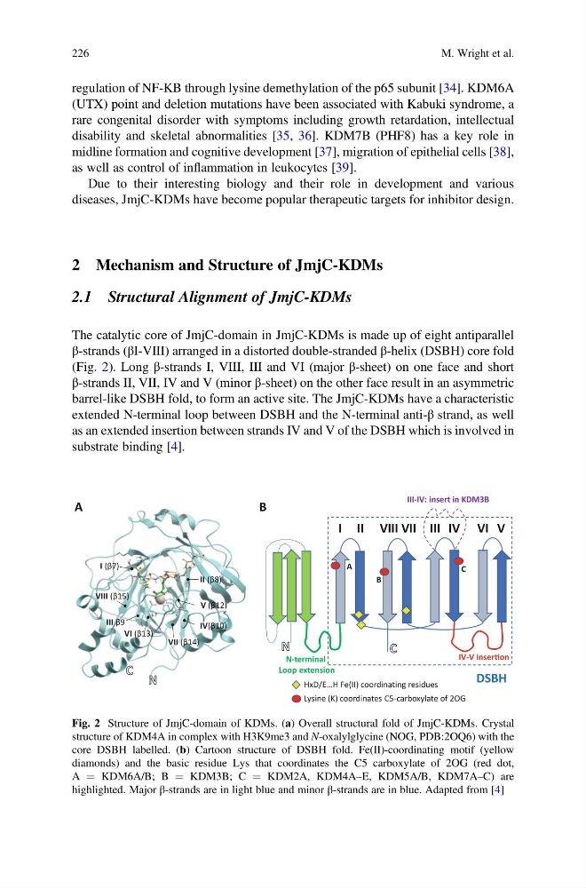
The catalytic core of JmjC-domain in JmjC-KDMs is made up of eight antiparallel
β-strands (βI-VIII) arranged in a distorted double-stranded β-helix (DSBH) core fold
(Fig. 2). Long β-strands I, VIII, III and VI (major β-sheet) on one face and short
β-strands II, VII, IV and V (minor β-sheet) on the other face result in an asymmetric
barrel-like DSBH fold, to form an active site. The JmjC-KDMs have a characteristic
extended N-terminal loop between DSBH and the N-terminal anti-β strand, as well
as an extended insertion between strands IV and V of the DSBH which is involved in
substrate binding [4].
I (β7)
VIII (β15)
III β9
VI (β13)
VII (β14)
V (β12)
IV(β10)
II (β8)
I II
III IV VI V
VIII VII
HxD/E…H Fe(II) coordinating residues
IV-V inserƟon
III-IV: insert in KDM3B
A
B
C
Lysine (K) coordinates C5-carboxylate of 2OG
A
B
DSBH
N-terminal
Loop extension
Fig. 2 Structure of JmjC-domain of KDMs. (a) Overall structural fold of JmjC-KDMs. Crystal
structure of KDM4A in complex with H3K9me3 and N-oxalylglycine (NOG, PDB:2OQ6) with the
core DSBH labelled. (b) Cartoon structure of DSBH fold. Fe(II)-coordinating motif (yellow
diamonds) and the basic residue Lys that coordinates the C5 carboxylate of 2OG (red dot,
A ¼ KDM6A/B; B ¼ KDM3B; C ¼ KDM2A, KDM4A–E, KDM5A/B, KDM7A–C) are
highlighted. Major β-strands are in light blue and minor β-strands are in blue. Adapted from [4]
226
M. Wright et al.
(UTX) point and deletion mutations have been associated with Kabuki syndrome, a
rare congenital disorder with symptoms including growth retardation, intellectual
disability and skeletal abnormalities [35, 36]. KDM7B (PHF8) has a key role in
midline formation and cognitive development [37], migration of epithelial cells [38],
as well as control of inflammation in leukocytes [39].
Due to their interesting biology and their role in development and various
diseases, JmjC-KDMs have become popular therapeutic targets for inhibitor design.
2 Mechanism and Structure of JmjC-KDMs
2.1 Structural Alignment of JmjC-KDMs
The catalytic core of JmjC-domain in JmjC-KDMs is made up of eight antiparallel
β-strands (βI-VIII) arranged in a distorted double-stranded β-helix (DSBH) core fold
(Fig. 2). Long β-strands I, VIII, III and VI (major β-sheet) on one face and short
β-strands II, VII, IV and V (minor β-sheet) on the other face result in an asymmetric
barrel-like DSBH fold, to form an active site. The JmjC-KDMs have a characteristic
extended N-terminal loop between DSBH and the N-terminal anti-β strand, as well
as an extended insertion between strands IV and V of the DSBH which is involved in
substrate binding [4].
I (β7)
VIII (β15)
III β9
VI (β13)
VII (β14)
V (β12)
IV(β10)
II (β8)
I II
III IV VI V
VIII VII
HxD/E…H Fe(II) coordinating residues
IV-V inserƟon
III-IV: insert in KDM3B
A
B
C
Lysine (K) coordinates C5-carboxylate of 2OG
A
B
DSBH
N-terminal
Loop extension
Fig. 2 Structure of JmjC-domain of KDMs. (a) Overall structural fold of JmjC-KDMs. Crystal
structure of KDM4A in complex with H3K9me3 and N-oxalylglycine (NOG, PDB:2OQ6) with the
core DSBH labelled. (b) Cartoon structure of DSBH fold. Fe(II)-coordinating motif (yellow
diamonds) and the basic residue Lys that coordinates the C5 carboxylate of 2OG (red dot,
A ¼ KDM6A/B; B ¼ KDM3B; C ¼ KDM2A, KDM4A–E, KDM5A/B, KDM7A–C) are
highlighted. Major β-strands are in light blue and minor β-strands are in blue. Adapted from [4]
226
M. Wright et al.