

2.1.1 2OG Cofactor Binding Site
The JmjC-KDMs have structurally similar active sites in line with their catalytically
similar mechanisms, where 2OG cofactor binds to the catalytic Fe(II) centre through
bidentate coordination of the keto acid (Fig. 1). In all KDM structures, the metal ion
is positioned through interaction with a triad of residues that forms a HxD/E . . . H
motif (Table 1). Note that catalytically inactive 2OG analogue, N-oxalylglycine
(NOG), and Ni(II) or Mn(II) are often used for JmjC-KDM crystallography. The
two His residues are positioned distally on βII, βVII or βVIII, and the Asp or Glu
residue is located on βII/βIII loop (Fig. 2). For KDM2A/B, KDM3A/B and KDM7A/
B, two histidines and an aspartic acid coordinate to the metal centre. For KDM4
(JMJD2A, B, C and D), KDM5 (JARID1A, B, C and D) and KDM6 (JMJD3, UTX
and UTY), the acidic aspartate residue is replaced with a glutamic acid. PHF2
(KDM7C) is anomalous with a histidine, aspartic acid and tyrosine residues coordinating to the metal centre. The three metal coordinating residues are essential for
catalysis, and Ala mutants have been shown to abolish the enzyme activity, and often
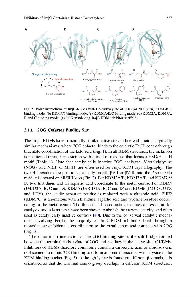
used as catalytically inactive controls [40]. Due to the conserved catalytic mechanism involving Fe(II), the majority of JmjC-KDM inhibitors bind through a
monodentate or bidentate coordination to the metal centre and compete with 2OG
(Fig. 3).
The other main interaction at the 2OG-binding site is the salt bridge formed
between the terminal carboxylate of 2OG and residues in the active site of KDMs.
Inhibitors of KDMs therefore commonly contain a carboxylic acid or a bioisosteric
replacement to mimic 2OG binding and form an ionic interaction with a lysine in the
KDM binding pocket (Fig. 3). Although lysine is found on different β-strands, it is
orientated so that the terminal amino group overlaps in different KDM structures.
Fig. 3 Polar interactions of JmjC-KDMs with C5-carboxylate of 2OG (or NOG): (a) KDM3B/C
binding mode; (b) KDM4/5 binding mode; (c) KDM6A/B/C binding mode; (d) KDM2A, KDM7A,
B and C binding mode; (e) 2OG mimicking JmjC-KDM inhibitor scaffolds
Inhibitors of JmjC-Containing Histone Demethylases
227
The JmjC-KDMs have structurally similar active sites in line with their catalytically
similar mechanisms, where 2OG cofactor binds to the catalytic Fe(II) centre through
bidentate coordination of the keto acid (Fig. 1). In all KDM structures, the metal ion
is positioned through interaction with a triad of residues that forms a HxD/E . . . H
motif (Table 1). Note that catalytically inactive 2OG analogue, N-oxalylglycine
(NOG), and Ni(II) or Mn(II) are often used for JmjC-KDM crystallography. The
two His residues are positioned distally on βII, βVII or βVIII, and the Asp or Glu
residue is located on βII/βIII loop (Fig. 2). For KDM2A/B, KDM3A/B and KDM7A/
B, two histidines and an aspartic acid coordinate to the metal centre. For KDM4
(JMJD2A, B, C and D), KDM5 (JARID1A, B, C and D) and KDM6 (JMJD3, UTX
and UTY), the acidic aspartate residue is replaced with a glutamic acid. PHF2
(KDM7C) is anomalous with a histidine, aspartic acid and tyrosine residues coordinating to the metal centre. The three metal coordinating residues are essential for
catalysis, and Ala mutants have been shown to abolish the enzyme activity, and often
used as catalytically inactive controls [40]. Due to the conserved catalytic mechanism involving Fe(II), the majority of JmjC-KDM inhibitors bind through a
monodentate or bidentate coordination to the metal centre and compete with 2OG
(Fig. 3).
The other main interaction at the 2OG-binding site is the salt bridge formed
between the terminal carboxylate of 2OG and residues in the active site of KDMs.
Inhibitors of KDMs therefore commonly contain a carboxylic acid or a bioisosteric
replacement to mimic 2OG binding and form an ionic interaction with a lysine in the
KDM binding pocket (Fig. 3). Although lysine is found on different β-strands, it is
orientated so that the terminal amino group overlaps in different KDM structures.
Fig. 3 Polar interactions of JmjC-KDMs with C5-carboxylate of 2OG (or NOG): (a) KDM3B/C
binding mode; (b) KDM4/5 binding mode; (c) KDM6A/B/C binding mode; (d) KDM2A, KDM7A,
B and C binding mode; (e) 2OG mimicking JmjC-KDM inhibitor scaffolds
Inhibitors of JmjC-Containing Histone Demethylases
227