

MiDAC, while HDAC3 is part of NcoR and SMRT complexes, and HDAC8 can
function independently of multiprotein complexes. Compared to class I, the class IIa
isoforms are tissue-specific, larger in size, and shuttle between the nucleus and
cytoplasm upon activation. The class IIa enzymes have weak deacetylase activity
in vitro, and relatively little is known about their substrates. It is possible that their
primary function is in fact not enzymatic and they function as a scaffold to bring
together multiprotein complexes [18]. The class IIa isoforms are found in NCoR and
SMRT protein complexes, and the region involved in protein-protein interaction
with the HDACs has been identified [19]. The class IIb isoforms HDAC6 and
HDAC10 are primarily cytoplasmic in location, as reflected in their known substrates. HDAC6 has two catalytic domains although clear catalytic activity was
demonstrated with only one of these [20]. Finally, HDAC11 is placed distinctly in
class IV as it has similarities to both the class I and class II isoforms [21]. Although
the 11 HDAC isoforms overlap with one another in terms of localization and
substrates, they clearly play nonredundant physiological roles as evidenced by the
fact that only HDAC6, HDAC10, and HDAC11 knockouts produce a viable phenotype in mice.
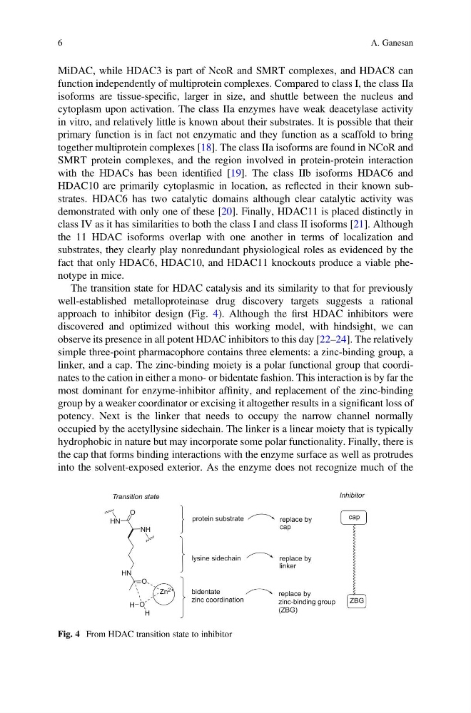
The transition state for HDAC catalysis and its similarity to that for previously
well-established metalloproteinase drug discovery targets suggests a rational
approach to inhibitor design (Fig. 4). Although the first HDAC inhibitors were
discovered and optimized without this working model, with hindsight, we can
observe its presence in all potent HDAC inhibitors to this day [22–24]. The relatively
simple three-point pharmacophore contains three elements: a zinc-binding group, a
linker, and a cap. The zinc-binding moiety is a polar functional group that coordinates to the cation in either a mono- or bidentate fashion. This interaction is by far the
most dominant for enzyme-inhibitor affinity, and replacement of the zinc-binding
group by a weaker coordinator or excising it altogether results in a significant loss of
potency. Next is the linker that needs to occupy the narrow channel normally
occupied by the acetyllysine sidechain. The linker is a linear moiety that is typically
hydrophobic in nature but may incorporate some polar functionality. Finally, there is
the cap that forms binding interactions with the enzyme surface as well as protrudes
into the solvent-exposed exterior. As the enzyme does not recognize much of the
NH
HN
O
HN
O
H
O
H
Zn 2+
Transition state
replace by
zinc-binding group
(ZBG)
replace by
linker
bidentate
zinc coordination
lysine sidechain
protein substrate
replace by
cap
Inhibitor
ZBG
cap
Fig. 4 From HDAC transition state to inhibitor
6
A. Ganesan
function independently of multiprotein complexes. Compared to class I, the class IIa
isoforms are tissue-specific, larger in size, and shuttle between the nucleus and
cytoplasm upon activation. The class IIa enzymes have weak deacetylase activity
in vitro, and relatively little is known about their substrates. It is possible that their
primary function is in fact not enzymatic and they function as a scaffold to bring
together multiprotein complexes [18]. The class IIa isoforms are found in NCoR and
SMRT protein complexes, and the region involved in protein-protein interaction
with the HDACs has been identified [19]. The class IIb isoforms HDAC6 and
HDAC10 are primarily cytoplasmic in location, as reflected in their known substrates. HDAC6 has two catalytic domains although clear catalytic activity was
demonstrated with only one of these [20]. Finally, HDAC11 is placed distinctly in
class IV as it has similarities to both the class I and class II isoforms [21]. Although
the 11 HDAC isoforms overlap with one another in terms of localization and
substrates, they clearly play nonredundant physiological roles as evidenced by the
fact that only HDAC6, HDAC10, and HDAC11 knockouts produce a viable phenotype in mice.
The transition state for HDAC catalysis and its similarity to that for previously
well-established metalloproteinase drug discovery targets suggests a rational
approach to inhibitor design (Fig. 4). Although the first HDAC inhibitors were
discovered and optimized without this working model, with hindsight, we can
observe its presence in all potent HDAC inhibitors to this day [22–24]. The relatively
simple three-point pharmacophore contains three elements: a zinc-binding group, a
linker, and a cap. The zinc-binding moiety is a polar functional group that coordinates to the cation in either a mono- or bidentate fashion. This interaction is by far the
most dominant for enzyme-inhibitor affinity, and replacement of the zinc-binding
group by a weaker coordinator or excising it altogether results in a significant loss of
potency. Next is the linker that needs to occupy the narrow channel normally
occupied by the acetyllysine sidechain. The linker is a linear moiety that is typically
hydrophobic in nature but may incorporate some polar functionality. Finally, there is
the cap that forms binding interactions with the enzyme surface as well as protrudes
into the solvent-exposed exterior. As the enzyme does not recognize much of the
NH
HN
O
HN
O
H
O
H
Zn 2+
Transition state
replace by
zinc-binding group
(ZBG)
replace by
linker
bidentate
zinc coordination
lysine sidechain
protein substrate
replace by
cap
Inhibitor
ZBG
cap
Fig. 4 From HDAC transition state to inhibitor
6
A. Ganesan