

glycosylated linker-peptide. However, by the enzymatic treatment with papain, it
can be departed easily into the two operative domains (Tomme et al. 1988)
(Fig. 11.2).
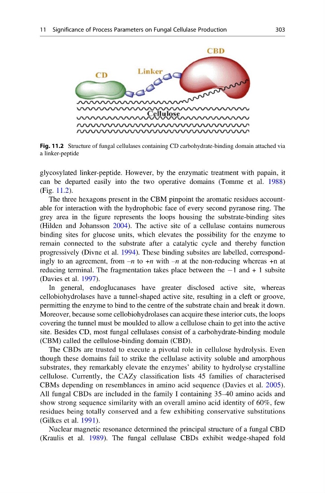
The three hexagons present in the CBM pinpoint the aromatic residues accountable for interaction with the hydrophobic face of every second pyranose ring. The
grey area in the figure represents the loops housing the substrate-binding sites
(Hilden and Johansson 2004). The active site of a cellulase contains numerous
binding sites for glucose units, which elevates the possibility for the enzyme to
remain connected to the substrate after a catalytic cycle and thereby function
progressively (Divne et al. 1994). These binding subsites are labelled, correspondingly to an agreement, from –n to +n with –n at the non-reducing whereas +n at
reducing terminal. The fragmentation takes place between the À1 and + 1 subsite
(Davies et al. 1997).
In general, endoglucanases have greater disclosed active site, whereas
cellobiohydrolases have a tunnel-shaped active site, resulting in a cleft or groove,
permitting the enzyme to bind to the centre of the substrate chain and break it down.
Moreover, because some cellobiohydrolases can acquire these interior cuts, the loops
covering the tunnel must be moulded to allow a cellulose chain to get into the active
site. Besides CD, most fungal cellulases consist of a carbohydrate-binding module
(CBM) called the cellulose-binding domain (CBD).
The CBDs are trusted to execute a pivotal role in cellulose hydrolysis. Even
though these domains fail to strike the cellulase activity soluble and amorphous
substrates, they remarkably elevate the enzymes’ ability to hydrolyse crystalline
cellulose. Currently, the CAZy classification lists 45 families of characterised
CBMs depending on resemblances in amino acid sequence (Davies et al. 2005).
All fungal CBDs are included in the family I containing 35–40 amino acids and
show strong sequence similarity with an overall amino acid identity of 60%, few
residues being totally conserved and a few exhibiting conservative substitutions
(Gilkes et al. 1991).
Nuclear magnetic resonance determined the principal structure of a fungal CBD
(Kraulis et al. 1989). The fungal cellulase CBDs exhibit wedge-shaped fold
Fig. 11.2 Structure of fungal cellulases containing CD carbohydrate-binding domain attached via
a linker-peptide
11 Significance of Process Parameters on Fungal Cellulase Production
303
can be departed easily into the two operative domains (Tomme et al. 1988)
(Fig. 11.2).
The three hexagons present in the CBM pinpoint the aromatic residues accountable for interaction with the hydrophobic face of every second pyranose ring. The
grey area in the figure represents the loops housing the substrate-binding sites
(Hilden and Johansson 2004). The active site of a cellulase contains numerous
binding sites for glucose units, which elevates the possibility for the enzyme to
remain connected to the substrate after a catalytic cycle and thereby function
progressively (Divne et al. 1994). These binding subsites are labelled, correspondingly to an agreement, from –n to +n with –n at the non-reducing whereas +n at
reducing terminal. The fragmentation takes place between the À1 and + 1 subsite
(Davies et al. 1997).
In general, endoglucanases have greater disclosed active site, whereas
cellobiohydrolases have a tunnel-shaped active site, resulting in a cleft or groove,
permitting the enzyme to bind to the centre of the substrate chain and break it down.
Moreover, because some cellobiohydrolases can acquire these interior cuts, the loops
covering the tunnel must be moulded to allow a cellulose chain to get into the active
site. Besides CD, most fungal cellulases consist of a carbohydrate-binding module
(CBM) called the cellulose-binding domain (CBD).
The CBDs are trusted to execute a pivotal role in cellulose hydrolysis. Even
though these domains fail to strike the cellulase activity soluble and amorphous
substrates, they remarkably elevate the enzymes’ ability to hydrolyse crystalline
cellulose. Currently, the CAZy classification lists 45 families of characterised
CBMs depending on resemblances in amino acid sequence (Davies et al. 2005).
All fungal CBDs are included in the family I containing 35–40 amino acids and
show strong sequence similarity with an overall amino acid identity of 60%, few
residues being totally conserved and a few exhibiting conservative substitutions
(Gilkes et al. 1991).
Nuclear magnetic resonance determined the principal structure of a fungal CBD
(Kraulis et al. 1989). The fungal cellulase CBDs exhibit wedge-shaped fold
Fig. 11.2 Structure of fungal cellulases containing CD carbohydrate-binding domain attached via
a linker-peptide
11 Significance of Process Parameters on Fungal Cellulase Production
303