

PLFAs with respect to bacterial groups, this method is nowadays widely used for
rapid quantification of microbial biomass in samples (e.g., in soil and sludge) with an
insight into the composition of basic bacterial groups. In this way, PLFA analysis
also enables to observe extensive changes in the community composition of the
living microbes in soil and water environments. Despite certain concerns about the
assignment of individual PLFA to bacterial taxonomic groups, some typical PLFAs
represent dominant groups of bacteria and in this way the biomass of these groups
can be quantified. The main advantage of the PLFA method is its simple use and
precise analytical protocols for determination of the monitored analytes without the
need for cultivation steps. Another important positive aspect is the fact that PLFAs
decompose relatively quickly via phosphate removal from the original structure, and,
therefore, PLFAs represent only the active and living biomass.
Typical protocol for the determination of PLFAs encompasses extraction of the
total lipids, separation of the PLFA fraction, derivatization (mostly methylation), and
analysis by gas chromatography. The total lipids from the samples are usually
extracted via the Bligh and Dyer method (Bligh and Dyer 1959) using a mixture
of chloroform, methanol, and phosphate buffer (1:2:0.8; v/v/v). Once the total lipids
are extracted, the samples are separated using a simple column chromatography with
available solid-phase extraction silica gel columns. Due to their polarity, PLFAs are
easily separated from other lipids. The samples are typically subjected to mild
alkaline methanolysis resulting in methyl esters of fatty acids originating from the
phospholipid fraction (Sampedro et al. 2009). Methyl esters of fatty acids are
routinely analyzed using high-resolution capillary gas chromatography with either
flame ionization detector or any hyphenated mass detector.
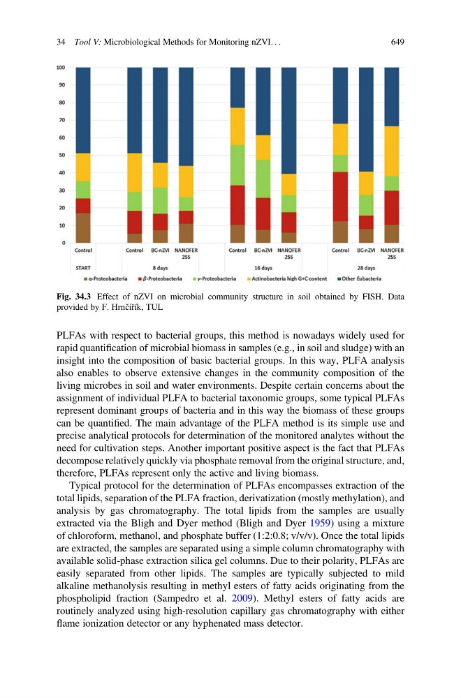
Fig. 34.3 Effect of nZVI on microbial community structure in soil obtained by FISH. Data
provided by F. Hrnčiřík, TUL
34 Tool V: Microbiological Methods for Monitoring nZVI. . .
649