

oligonucleotide probes; however, the specificity is lower. The sensitivity of FISH in
natural water or soil samples considerably increased when combined with catalyzed
reporter deposition (CARD)-FISH (Kubota 2013). Recently direct-geneFISH, which
allows a direct cell identification based on rRNA-targeted oligonucleotide probes
and gene detection with dsDNA polynucleotide probes, has been developed for
environmental samples (Barrero-Canosa et al. 2017). The advantage is in a single
hybridization, taking far less time than CARD-FISH.
Here, a study on the nZVI effect on natural soil bacteria using FISH is described
as an example. First, soil mesocosms were prepared with 5% of nZVI materials
(NANOFER 25S and biochar-embedded (BC)-nZVI). After incubation, the bacteria
were separated from soil using density gradient centrifugation, filtrated, and hybridized with specific probes for Eubacteria (EUB338 probe), Alfaproteobacteria
(ALF1B), Betaproteobacteria (BET42a), Gammaproteobacteria (GAM42a), and
Actinobacteria (HGC69A). After the hybridization, each filter was mounted using
antifadent Citifluor solution on a glass slide and typically ten images for each probe
were taken using fluorescence Axio Imager microscope and AxioVision software
(Zeiss). The evaluation of the FISH images was then performed in Matlab (The
Mathworks) with Image Processing Toolbox. The results were finally expressed as
relative abundance of the studied bacterial groups (Fig. 34.3).
34.4 Phospholipid Fatty Acid (PLFA) Analysis
PLFA analysis represents another suitable method for studying microorganisms in
environmental samples. The principle lies in a quantitative analysis of fatty acids
originating from phospholipids, which are an essential structural component of all
microbial cellular membranes. However, this application is shadowed by a rapid
development and availability of more effective molecular biology methods, which
are also described in this chapter. On the other hand, due to certain specificity of
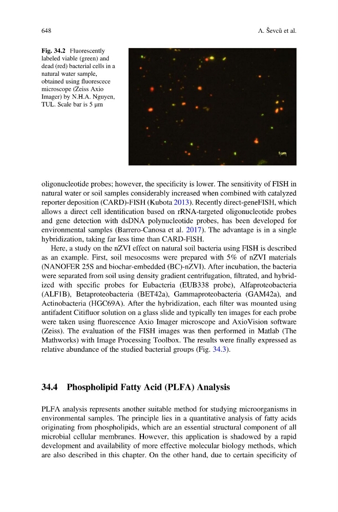
Fig. 34.2 Fluorescently
labeled viable (green) and
dead (red) bacterial cells in a
natural water sample,
obtained using fluorescece
microscope (Zeiss Axio
Imager) by N.H.A. Nguyen,
TUL. Scale bar is 5 μm
648
A. Ševců et al.