

The membrane filter method is often used to estimate the CFU counts from
samples with low bacterial numbers. A known volume of sample is filtered through
a membrane filter, then the filter is placed onto a Petri dish with a growth medium.
The nutrients diffuse through the filter from the medium and enable the development
of colonies. After an adequate incubation period, the CFU count on the surface of the
filter can be determined.
34.3 Fluorescence Microscopy
Fluorescence microscopy techniques can be used in order to examine the effect of
nZVI on organisms living in soil or groundwater environments. Such techniques
include fluorescence staining for total counts, viability counts, and fluorescence in
situ hybridization (FISH). For instance, the total cell number, e.g., expressed as the
number of bacteria per mL or L in liquid samples or per g of dry weight in soil
samples, can be determined by direct counting using DAPI (4
0 ,6-diamidino-2phenylindole) staining as a DNA fluorescence agent.
Cell viability, besides other methods, is assessed using a two-dye fluorescent
bacterial viability kit that distinguishes viable (commonly green fluorescence) and
dead (red fluorescence) cells under a fluorescence microscope (Fig. 34.2). Afterwards, it is possible to calculate the ratio of live and dead cells in the sample.
The composition of the soil or groundwater microbial communities is often
analyzed by FISH. A key advantage of this technique is the ability to observe
individual cells using fluorescence microscopy or flow cytometry. Thus, it can
give detailed information about morphology, number, and spatial distribution of
the targeted microbial groups. In brief, FISH detects nucleic acid sequences using
fluorescently labeled probe that hybridizes specifically to its complementary target
sequence (in rRNA, mRNA, and functional genes). The choice of the probes has to
be based on specificity, sensitivity, and ease of cell penetration. The most common
and commercially available are single-molecule-labeled oligonucleotide probes. A
polynucleotide probe has several fluorophores, resulting in higher sensitivity than
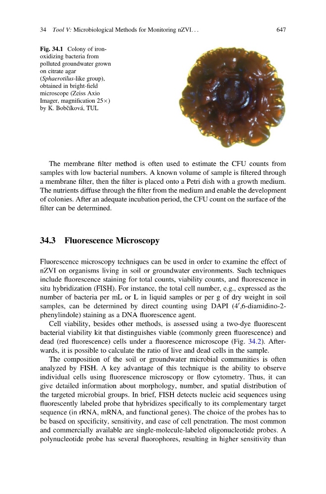
Fig. 34.1 Colony of ironoxidizing bacteria from
polluted groundwater grown
on citrate agar
(Sphaerotilus-like group),
obtained in bright-field
microscope (Zeiss Axio
Imager, magnification 25Â)
by K. Bobčíková, TUL
34 Tool V: Microbiological Methods for Monitoring nZVI. . .
647