

396
Y. Dong et al.
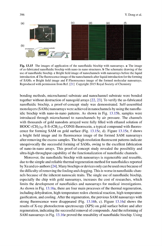
Fig. 13.15 The images of application of the nanofluidic biochip with nanoarrays. a The image
of as-fabricated nanofluidic biochip with nano-in-nano structures. b The schematic drawing of the
use of nanofluidic biochip. c Bright field image of nanochannels with nanoarrays before the liquid
introduction. d The fluorescence image of the nanochannels after liquid introduction for the forming
of SAMs. e Bright field image and f Fluorescence image of the formed molecular nanoarrays.
Reproduced with permission from Ref. [21] Copyright 2015 Royal Society of Chemistry
bonding methods, microchannel substrate and nanochannel substrate were bonded
together without destruction of nanogold arrays [22, 23]. To verify the as-fabricated
nanofluidic biochip, a proof-of-concept study was demonstrated. Self-assembled
monolayers (SAMs) nanoarrays were achieved in nanochannels by using the nanofluidic biochip with nano-in-nano patterns. As shown in Fig. 13.15b, samples were
introduced through microchannel to nanochannels by air pressure. The channels
with thousands of gold nanodots arrayed were fully filled with ethanol solution of
HOOC-(CH 2 ) 10 -S-S-(CH 2 ) 10 -CONH-fluorescein, a typical compound with fluorescence for forming SAM on gold surface (Fig. 13.15c, d). Figure 13.15e, f shows
a bright field image and its fluorescence image of the formed SAM nanoarrays
after removing the excess samples. The high-resolution fluorescent patterns indicate
unequivocally the successful forming of SAMs, owing to the excellent fabrication
of nano-in-nano arrays. This proof-of-concept study revealed the possibility and
ultra-high-throughput capability of the functionalization of nanofluidic channels.
Moreover, the nanofluidic biochip with nanoarrays is regenerable and reusable,
due to the simple and reliable thermal regeneration method for nanofluidics reported
by Xu and co-authors [24]. Many biochips or devices only can be used once because of
the difficulty of removing the fouling and clogging. This is worse in nanofluidic channels because of the inherent nanoscale traits. The single use of nanofluidic biochip,
especially the chip with gold nanoarrays, increases the cost of researches, which
limits the development of nanofluidics and nanoarrays for medical investigations.
As shown in Fig. 13.16a, there are four main processes of the thermal regeneration,
including dehydration, high-temperature redox chemical reaction, high-temperature
gasification, and cooling. After the regeneration, the previous SAM nanoarrays with
strong fluorescence were disappeared (Fig. 13.16b, c). Figure 13.16d shows the
results of X-ray photoelectron spectroscopy (XPS) on gold surface before and after
regeneration, indicating the successful removal of compounds. And the reforming of
SAM nanoarrays in Fig. 13.16e proved the reusability of nanofluidic biochip. Using
Y. Dong et al.
Fig. 13.15 The images of application of the nanofluidic biochip with nanoarrays. a The image
of as-fabricated nanofluidic biochip with nano-in-nano structures. b The schematic drawing of the
use of nanofluidic biochip. c Bright field image of nanochannels with nanoarrays before the liquid
introduction. d The fluorescence image of the nanochannels after liquid introduction for the forming
of SAMs. e Bright field image and f Fluorescence image of the formed molecular nanoarrays.
Reproduced with permission from Ref. [21] Copyright 2015 Royal Society of Chemistry
bonding methods, microchannel substrate and nanochannel substrate were bonded
together without destruction of nanogold arrays [22, 23]. To verify the as-fabricated
nanofluidic biochip, a proof-of-concept study was demonstrated. Self-assembled
monolayers (SAMs) nanoarrays were achieved in nanochannels by using the nanofluidic biochip with nano-in-nano patterns. As shown in Fig. 13.15b, samples were
introduced through microchannel to nanochannels by air pressure. The channels
with thousands of gold nanodots arrayed were fully filled with ethanol solution of
HOOC-(CH 2 ) 10 -S-S-(CH 2 ) 10 -CONH-fluorescein, a typical compound with fluorescence for forming SAM on gold surface (Fig. 13.15c, d). Figure 13.15e, f shows
a bright field image and its fluorescence image of the formed SAM nanoarrays
after removing the excess samples. The high-resolution fluorescent patterns indicate
unequivocally the successful forming of SAMs, owing to the excellent fabrication
of nano-in-nano arrays. This proof-of-concept study revealed the possibility and
ultra-high-throughput capability of the functionalization of nanofluidic channels.
Moreover, the nanofluidic biochip with nanoarrays is regenerable and reusable,
due to the simple and reliable thermal regeneration method for nanofluidics reported
by Xu and co-authors [24]. Many biochips or devices only can be used once because of
the difficulty of removing the fouling and clogging. This is worse in nanofluidic channels because of the inherent nanoscale traits. The single use of nanofluidic biochip,
especially the chip with gold nanoarrays, increases the cost of researches, which
limits the development of nanofluidics and nanoarrays for medical investigations.
As shown in Fig. 13.16a, there are four main processes of the thermal regeneration,
including dehydration, high-temperature redox chemical reaction, high-temperature
gasification, and cooling. After the regeneration, the previous SAM nanoarrays with
strong fluorescence were disappeared (Fig. 13.16b, c). Figure 13.16d shows the
results of X-ray photoelectron spectroscopy (XPS) on gold surface before and after
regeneration, indicating the successful removal of compounds. And the reforming of
SAM nanoarrays in Fig. 13.16e proved the reusability of nanofluidic biochip. Using