

13 Some Frontier Technologies for Aptamers in Medical Applications
393
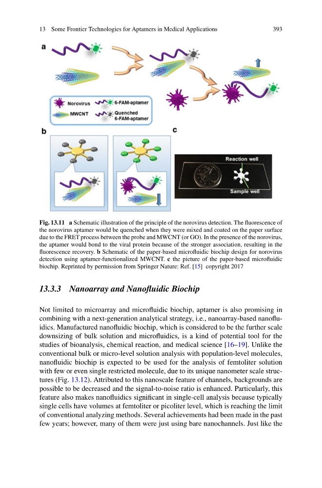
Fig. 13.11 a Schematic illustration of the principle of the norovirus detection. The fluorescence of
the norovirus aptamer would be quenched when they were mixed and coated on the paper surface
due to the FRET process between the probe and MWCNT (or GO). In the presence of the norovirus,
the aptamer would bond to the viral protein because of the stronger association, resulting in the
fluorescence recovery. b Schematic of the paper-based microfluidic biochip design for norovirus
detection using aptamer-functionalized MWCNT. c the picture of the paper-based microfluidic
biochip. Reprinted by permission from Springer Nature: Ref. [15] copyright 2017
13.3.3 Nanoarray and Nanofluidic Biochip
Not limited to microarray and microfluidic biochip, aptamer is also promising in
combining with a next-generation analytical strategy, i.e., nanoarray-based nanofluidics. Manufactured nanofluidic biochip, which is considered to be the further scale
downsizing of bulk solution and microfluidics, is a kind of potential tool for the
studies of bioanalysis, chemical reaction, and medical science [16–19]. Unlike the
conventional bulk or micro-level solution analysis with population-level molecules,
nanofluidic biochip is expected to be used for the analysis of femtoliter solution
with few or even single restricted molecule, due to its unique nanometer scale structures (Fig. 13.12). Attributed to this nanoscale feature of channels, backgrounds are
possible to be decreased and the signal-to-noise ratio is enhanced. Particularly, this
feature also makes nanofluidics significant in single-cell analysis because typically
single cells have volumes at femtoliter or picoliter level, which is reaching the limit
of conventional analyzing methods. Several achievements had been made in the past
few years; however, many of them were just using bare nanochannels. Just like the
393
Fig. 13.11 a Schematic illustration of the principle of the norovirus detection. The fluorescence of
the norovirus aptamer would be quenched when they were mixed and coated on the paper surface
due to the FRET process between the probe and MWCNT (or GO). In the presence of the norovirus,
the aptamer would bond to the viral protein because of the stronger association, resulting in the
fluorescence recovery. b Schematic of the paper-based microfluidic biochip design for norovirus
detection using aptamer-functionalized MWCNT. c the picture of the paper-based microfluidic
biochip. Reprinted by permission from Springer Nature: Ref. [15] copyright 2017
13.3.3 Nanoarray and Nanofluidic Biochip
Not limited to microarray and microfluidic biochip, aptamer is also promising in
combining with a next-generation analytical strategy, i.e., nanoarray-based nanofluidics. Manufactured nanofluidic biochip, which is considered to be the further scale
downsizing of bulk solution and microfluidics, is a kind of potential tool for the
studies of bioanalysis, chemical reaction, and medical science [16–19]. Unlike the
conventional bulk or micro-level solution analysis with population-level molecules,
nanofluidic biochip is expected to be used for the analysis of femtoliter solution
with few or even single restricted molecule, due to its unique nanometer scale structures (Fig. 13.12). Attributed to this nanoscale feature of channels, backgrounds are
possible to be decreased and the signal-to-noise ratio is enhanced. Particularly, this
feature also makes nanofluidics significant in single-cell analysis because typically
single cells have volumes at femtoliter or picoliter level, which is reaching the limit
of conventional analyzing methods. Several achievements had been made in the past
few years; however, many of them were just using bare nanochannels. Just like the