

392
Y. Dong et al.
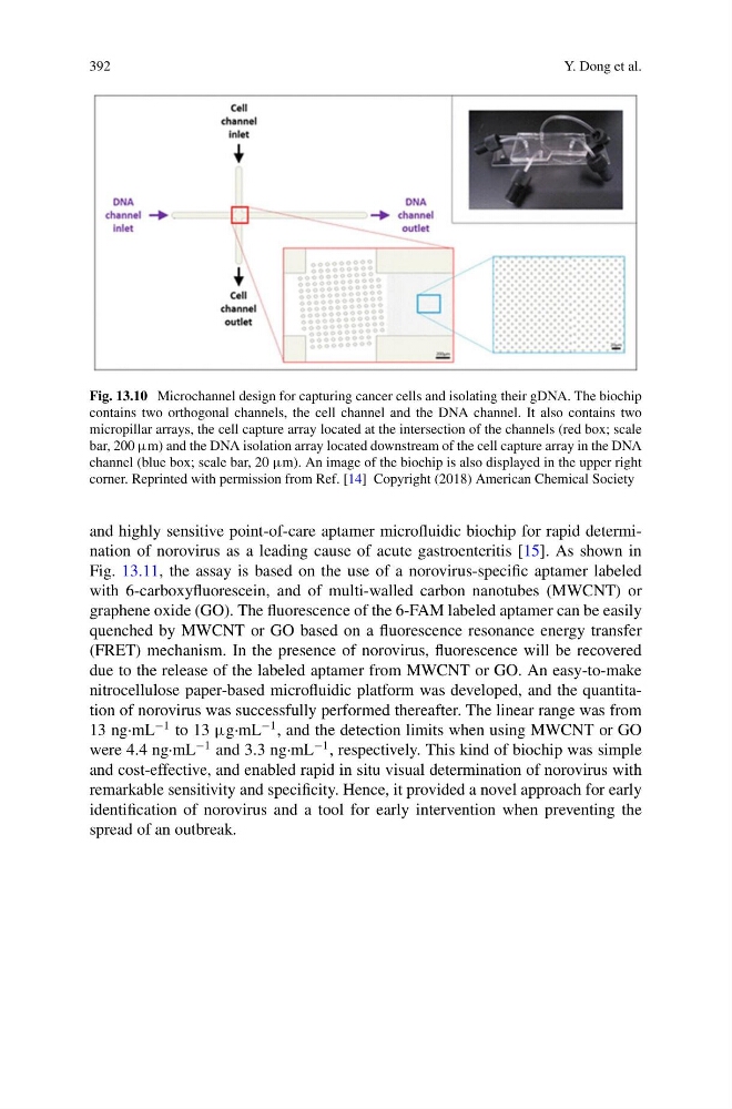
Fig. 13.10 Microchannel design for capturing cancer cells and isolating their gDNA. The biochip
contains two orthogonal channels, the cell channel and the DNA channel. It also contains two
micropillar arrays, the cell capture array located at the intersection of the channels (red box; scale
bar, 200 µm) and the DNA isolation array located downstream of the cell capture array in the DNA
channel (blue box; scale bar, 20 µm). An image of the biochip is also displayed in the upper right
corner. Reprinted with permission from Ref. [14] Copyright (2018) American Chemical Society
and highly sensitive point-of-care aptamer microfluidic biochip for rapid determination of norovirus as a leading cause of acute gastroenteritis [15]. As shown in
Fig. 13.11, the assay is based on the use of a norovirus-specific aptamer labeled
with 6-carboxyfluorescein, and of multi-walled carbon nanotubes (MWCNT) or
graphene oxide (GO). The fluorescence of the 6-FAM labeled aptamer can be easily
quenched by MWCNT or GO based on a fluorescence resonance energy transfer
(FRET) mechanism. In the presence of norovirus, fluorescence will be recovered
due to the release of the labeled aptamer from MWCNT or GO. An easy-to-make
nitrocellulose paper-based microfluidic platform was developed, and the quantitation of norovirus was successfully performed thereafter. The linear range was from
13 ng·mL
−1 to 13 µg·mL
−1 , and the detection limits when using MWCNT or GO
were 4.4 ng·mL
−1 and 3.3 ng·mL
−1 , respectively. This kind of biochip was simple
and cost-effective, and enabled rapid in situ visual determination of norovirus with
remarkable sensitivity and specificity. Hence, it provided a novel approach for early
identification of norovirus and a tool for early intervention when preventing the
spread of an outbreak.
Y. Dong et al.
Fig. 13.10 Microchannel design for capturing cancer cells and isolating their gDNA. The biochip
contains two orthogonal channels, the cell channel and the DNA channel. It also contains two
micropillar arrays, the cell capture array located at the intersection of the channels (red box; scale
bar, 200 µm) and the DNA isolation array located downstream of the cell capture array in the DNA
channel (blue box; scale bar, 20 µm). An image of the biochip is also displayed in the upper right
corner. Reprinted with permission from Ref. [14] Copyright (2018) American Chemical Society
and highly sensitive point-of-care aptamer microfluidic biochip for rapid determination of norovirus as a leading cause of acute gastroenteritis [15]. As shown in
Fig. 13.11, the assay is based on the use of a norovirus-specific aptamer labeled
with 6-carboxyfluorescein, and of multi-walled carbon nanotubes (MWCNT) or
graphene oxide (GO). The fluorescence of the 6-FAM labeled aptamer can be easily
quenched by MWCNT or GO based on a fluorescence resonance energy transfer
(FRET) mechanism. In the presence of norovirus, fluorescence will be recovered
due to the release of the labeled aptamer from MWCNT or GO. An easy-to-make
nitrocellulose paper-based microfluidic platform was developed, and the quantitation of norovirus was successfully performed thereafter. The linear range was from
13 ng·mL
−1 to 13 µg·mL
−1 , and the detection limits when using MWCNT or GO
were 4.4 ng·mL
−1 and 3.3 ng·mL
−1 , respectively. This kind of biochip was simple
and cost-effective, and enabled rapid in situ visual determination of norovirus with
remarkable sensitivity and specificity. Hence, it provided a novel approach for early
identification of norovirus and a tool for early intervention when preventing the
spread of an outbreak.