

13 Some Frontier Technologies for Aptamers in Medical Applications
391
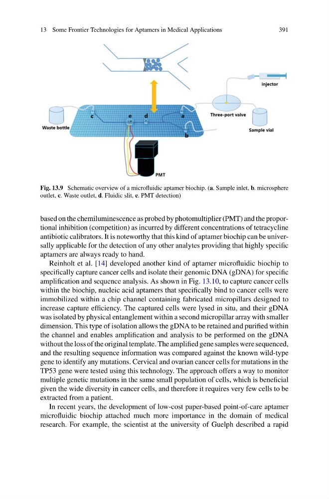
Fig. 13.9 Schematic overview of a microfluidic aptamer biochip. (a. Sample inlet, b. microsphere
outlet, c. Waste outlet, d. Fluidic slit, e. PMT detection)
based on the chemiluminescence as probed by photomultiplier (PMT) and the proportional inhibition (competition) as incurred by different concentrations of tetracycline
antibiotic calibrators. It is noteworthy that this kind of aptamer biochip can be universally applicable for the detection of any other analytes providing that highly specific
aptamers are always ready to hand.
Reinholt et al. [14] developed another kind of aptamer microfluidic biochip to
specifically capture cancer cells and isolate their genomic DNA (gDNA) for specific
amplification and sequence analysis. As shown in Fig. 13.10, to capture cancer cells
within the biochip, nucleic acid aptamers that specifically bind to cancer cells were
immobilized within a chip channel containing fabricated micropillars designed to
increase capture efficiency. The captured cells were lysed in situ, and their gDNA
was isolated by physical entanglement within a second micropillar array with smaller
dimension. This type of isolation allows the gDNA to be retained and purified within
the channel and enables amplification and analysis to be performed on the gDNA
without the loss of the original template. The amplified gene samples were sequenced,
and the resulting sequence information was compared against the known wild-type
gene to identify any mutations. Cervical and ovarian cancer cells for mutations in the
TP53 gene were tested using this technology. The approach offers a way to monitor
multiple genetic mutations in the same small population of cells, which is beneficial
given the wide diversity in cancer cells, and therefore it requires very few cells to be
extracted from a patient.
In recent years, the development of low-cost paper-based point-of-care aptamer
microfluidic biochip attached much more importance in the domain of medical
research. For example, the scientist at the university of Guelph described a rapid
391
Fig. 13.9 Schematic overview of a microfluidic aptamer biochip. (a. Sample inlet, b. microsphere
outlet, c. Waste outlet, d. Fluidic slit, e. PMT detection)
based on the chemiluminescence as probed by photomultiplier (PMT) and the proportional inhibition (competition) as incurred by different concentrations of tetracycline
antibiotic calibrators. It is noteworthy that this kind of aptamer biochip can be universally applicable for the detection of any other analytes providing that highly specific
aptamers are always ready to hand.
Reinholt et al. [14] developed another kind of aptamer microfluidic biochip to
specifically capture cancer cells and isolate their genomic DNA (gDNA) for specific
amplification and sequence analysis. As shown in Fig. 13.10, to capture cancer cells
within the biochip, nucleic acid aptamers that specifically bind to cancer cells were
immobilized within a chip channel containing fabricated micropillars designed to
increase capture efficiency. The captured cells were lysed in situ, and their gDNA
was isolated by physical entanglement within a second micropillar array with smaller
dimension. This type of isolation allows the gDNA to be retained and purified within
the channel and enables amplification and analysis to be performed on the gDNA
without the loss of the original template. The amplified gene samples were sequenced,
and the resulting sequence information was compared against the known wild-type
gene to identify any mutations. Cervical and ovarian cancer cells for mutations in the
TP53 gene were tested using this technology. The approach offers a way to monitor
multiple genetic mutations in the same small population of cells, which is beneficial
given the wide diversity in cancer cells, and therefore it requires very few cells to be
extracted from a patient.
In recent years, the development of low-cost paper-based point-of-care aptamer
microfluidic biochip attached much more importance in the domain of medical
research. For example, the scientist at the university of Guelph described a rapid