

3 Label-Free, Ultrahigh-Speed, Direct Imaging and Tracking …
71
term. In such interferometric detection, the scattering field of a nanoparticle and
the non-scattered transmitted field are often referred to as the signal and reference,
respectively. Notably, the phase difference ϕ between the scattering signal and the
transmitted reference is a function of the axial position of the particle relative to
the imaging microscope objective. This is because the phases of these two fields
evolve differently throughout the microscope objective due to their distinct spatial
modes [37]. In general, ϕ goes from 0 to π over the depth of focus of the imaging
microscope objective, which is approximately 1 μm for visible light and a high-NA
objective. In other words, ϕ can be manipulated by controlling the axial position
of the particle relative to the optical focus. In 2D imaging and tracking, the axial
position of the nanoparticle is adjusted by a sample stage to ensure that its absolute
contrast is maximized (as a bright spot when ϕ ∼ = 0 and as a dark spot when ϕ ∼ = π ).
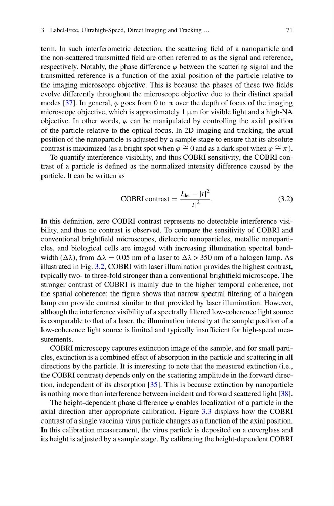
To quantify interference visibility, and thus COBRI sensitivity, the COBRI contrast of a particle is defined as the normalized intensity difference caused by the
particle. It can be written as
COBRI contrast =
I det − |t|
2
|t|
2
.
(3.2)
In this definition, zero COBRI contrast represents no detectable interference visibility, and thus no contrast is observed. To compare the sensitivity of COBRI and
conventional brightfield microscopes, dielectric nanoparticles, metallic nanoparticles, and biological cells are imaged with increasing illumination spectral bandwidth (λ), from λ = 0.05 nm of a laser to λ > 350 nm of a halogen lamp. As
illustrated in Fig. 3.2, COBRI with laser illumination provides the highest contrast,
typically two- to three-fold stronger than a conventional brightfield microscope. The
stronger contrast of COBRI is mainly due to the higher temporal coherence, not
the spatial coherence; the figure shows that narrow spectral filtering of a halogen
lamp can provide contrast similar to that provided by laser illumination. However,
although the interference visibility of a spectrally filtered low-coherence light source
is comparable to that of a laser, the illumination intensity at the sample position of a
low-coherence light source is limited and typically insufficient for high-speed measurements.
COBRI microscopy captures extinction image of the sample, and for small particles, extinction is a combined effect of absorption in the particle and scattering in all
directions by the particle. It is interesting to note that the measured extinction (i.e.,
the COBRI contrast) depends only on the scattering amplitude in the forward direction, independent of its absorption [35]. This is because extinction by nanoparticle
is nothing more than interference between incident and forward scattered light [38].
The height-dependent phase difference ϕ enables localization of a particle in the
axial direction after appropriate calibration. Figure 3.3 displays how the COBRI
contrast of a single vaccinia virus particle changes as a function of the axial position.
In this calibration measurement, the virus particle is deposited on a coverglass and
its height is adjusted by a sample stage. By calibrating the height-dependent COBRI
71
term. In such interferometric detection, the scattering field of a nanoparticle and
the non-scattered transmitted field are often referred to as the signal and reference,
respectively. Notably, the phase difference ϕ between the scattering signal and the
transmitted reference is a function of the axial position of the particle relative to
the imaging microscope objective. This is because the phases of these two fields
evolve differently throughout the microscope objective due to their distinct spatial
modes [37]. In general, ϕ goes from 0 to π over the depth of focus of the imaging
microscope objective, which is approximately 1 μm for visible light and a high-NA
objective. In other words, ϕ can be manipulated by controlling the axial position
of the particle relative to the optical focus. In 2D imaging and tracking, the axial
position of the nanoparticle is adjusted by a sample stage to ensure that its absolute
contrast is maximized (as a bright spot when ϕ ∼ = 0 and as a dark spot when ϕ ∼ = π ).
To quantify interference visibility, and thus COBRI sensitivity, the COBRI contrast of a particle is defined as the normalized intensity difference caused by the
particle. It can be written as
COBRI contrast =
I det − |t|
2
|t|
2
.
(3.2)
In this definition, zero COBRI contrast represents no detectable interference visibility, and thus no contrast is observed. To compare the sensitivity of COBRI and
conventional brightfield microscopes, dielectric nanoparticles, metallic nanoparticles, and biological cells are imaged with increasing illumination spectral bandwidth (λ), from λ = 0.05 nm of a laser to λ > 350 nm of a halogen lamp. As
illustrated in Fig. 3.2, COBRI with laser illumination provides the highest contrast,
typically two- to three-fold stronger than a conventional brightfield microscope. The
stronger contrast of COBRI is mainly due to the higher temporal coherence, not
the spatial coherence; the figure shows that narrow spectral filtering of a halogen
lamp can provide contrast similar to that provided by laser illumination. However,
although the interference visibility of a spectrally filtered low-coherence light source
is comparable to that of a laser, the illumination intensity at the sample position of a
low-coherence light source is limited and typically insufficient for high-speed measurements.
COBRI microscopy captures extinction image of the sample, and for small particles, extinction is a combined effect of absorption in the particle and scattering in all
directions by the particle. It is interesting to note that the measured extinction (i.e.,
the COBRI contrast) depends only on the scattering amplitude in the forward direction, independent of its absorption [35]. This is because extinction by nanoparticle
is nothing more than interference between incident and forward scattered light [38].
The height-dependent phase difference ϕ enables localization of a particle in the
axial direction after appropriate calibration. Figure 3.3 displays how the COBRI
contrast of a single vaccinia virus particle changes as a function of the axial position.
In this calibration measurement, the virus particle is deposited on a coverglass and
its height is adjusted by a sample stage. By calibrating the height-dependent COBRI