

70
C.-L. Hsieh
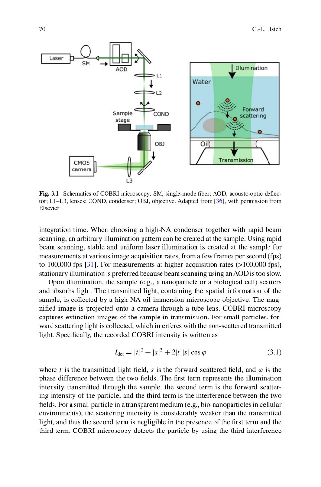
Fig. 3.1 Schematics of COBRI microscopy. SM, single-mode fiber; AOD, acousto-optic deflector; L1–L3, lenses; COND, condenser; OBJ, objective. Adapted from [36], with permission from
Elsevier
integration time. When choosing a high-NA condenser together with rapid beam
scanning, an arbitrary illumination pattern can be created at the sample. Using rapid
beam scanning, stable and uniform laser illumination is created at the sample for
measurements at various image acquisition rates, from a few frames per second (fps)
to 100,000 fps [31]. For measurements at higher acquisition rates (>100,000 fps),
stationary illumination is preferred because beam scanning using an AOD is too slow.
Upon illumination, the sample (e.g., a nanoparticle or a biological cell) scatters
and absorbs light. The transmitted light, containing the spatial information of the
sample, is collected by a high-NA oil-immersion microscope objective. The magnified image is projected onto a camera through a tube lens. COBRI microscopy
captures extinction images of the sample in transmission. For small particles, forward scattering light is collected, which interferes with the non-scattered transmitted
light. Specifically, the recorded COBRI intensity is written as
I det = |t|
2
+ |s|
2
+ 2|t||s| cos ϕ
(3.1)
where t is the transmitted light field, s is the forward scattered field, and ϕ is the
phase difference between the two fields. The first term represents the illumination
intensity transmitted through the sample; the second term is the forward scattering intensity of the particle, and the third term is the interference between the two
fields. For a small particle in a transparent medium (e.g., bio-nanoparticles in cellular
environments), the scattering intensity is considerably weaker than the transmitted
light, and thus the second term is negligible in the presence of the first term and the
third term. COBRI microscopy detects the particle by using the third interference
C.-L. Hsieh
Fig. 3.1 Schematics of COBRI microscopy. SM, single-mode fiber; AOD, acousto-optic deflector; L1–L3, lenses; COND, condenser; OBJ, objective. Adapted from [36], with permission from
Elsevier
integration time. When choosing a high-NA condenser together with rapid beam
scanning, an arbitrary illumination pattern can be created at the sample. Using rapid
beam scanning, stable and uniform laser illumination is created at the sample for
measurements at various image acquisition rates, from a few frames per second (fps)
to 100,000 fps [31]. For measurements at higher acquisition rates (>100,000 fps),
stationary illumination is preferred because beam scanning using an AOD is too slow.
Upon illumination, the sample (e.g., a nanoparticle or a biological cell) scatters
and absorbs light. The transmitted light, containing the spatial information of the
sample, is collected by a high-NA oil-immersion microscope objective. The magnified image is projected onto a camera through a tube lens. COBRI microscopy
captures extinction images of the sample in transmission. For small particles, forward scattering light is collected, which interferes with the non-scattered transmitted
light. Specifically, the recorded COBRI intensity is written as
I det = |t|
2
+ |s|
2
+ 2|t||s| cos ϕ
(3.1)
where t is the transmitted light field, s is the forward scattered field, and ϕ is the
phase difference between the two fields. The first term represents the illumination
intensity transmitted through the sample; the second term is the forward scattering intensity of the particle, and the third term is the interference between the two
fields. For a small particle in a transparent medium (e.g., bio-nanoparticles in cellular
environments), the scattering intensity is considerably weaker than the transmitted
light, and thus the second term is negligible in the presence of the first term and the
third term. COBRI microscopy detects the particle by using the third interference