

2 Interferometric Scattering (iSCAT) Microscopy and Related Techniques
45
sion of noise to the level of parts-per-million in order to overcome the challenge of
detecting a single molecule in extinction. While several groups attempted absorption
measurements in various configurations [154–156], it was iSCAT that demonstrated
direct room temperature modulation-free extinction of single molecules and quantum
dots [114, 133, 143].
The first important step in extending the sensitivity of iSCAT was taken with the
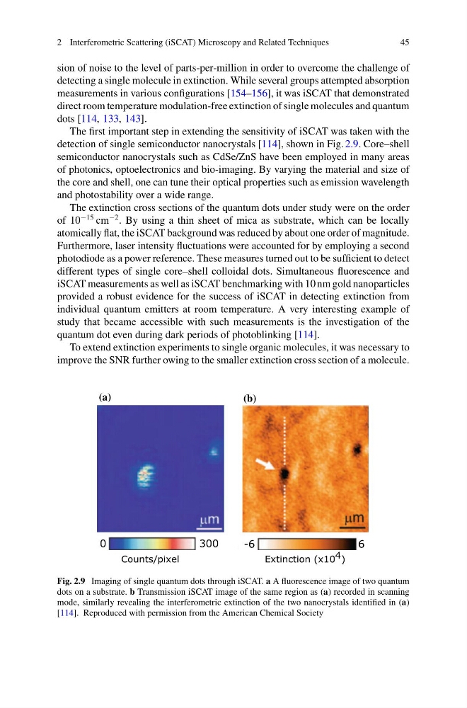
detection of single semiconductor nanocrystals [114], shown in Fig. 2.9. Core–shell
semiconductor nanocrystals such as CdSe/ZnS have been employed in many areas
of photonics, optoelectronics and bio-imaging. By varying the material and size of
the core and shell, one can tune their optical properties such as emission wavelength
and photostability over a wide range.
The extinction cross sections of the quantum dots under study were on the order
of 10
−15 cm
−2 . By using a thin sheet of mica as substrate, which can be locally
atomically flat, the iSCAT background was reduced by about one order of magnitude.
Furthermore, laser intensity fluctuations were accounted for by employing a second
photodiode as a power reference. These measures turned out to be sufficient to detect
different types of single core–shell colloidal dots. Simultaneous fluorescence and
iSCAT measurements as well as iSCAT benchmarking with 10 nm gold nanoparticles
provided a robust evidence for the success of iSCAT in detecting extinction from
individual quantum emitters at room temperature. A very interesting example of
study that became accessible with such measurements is the investigation of the
quantum dot even during dark periods of photoblinking [114].
To extend extinction experiments to single organic molecules, it was necessary to
improve the SNR further owing to the smaller extinction cross section of a molecule.
300
0
Counts/pixel
(a)
(b)
6
-6
Extinction (x10 4 )
Fig. 2.9 Imaging of single quantum dots through iSCAT. a A fluorescence image of two quantum
dots on a substrate. b Transmission iSCAT image of the same region as (a) recorded in scanning
mode, similarly revealing the interferometric extinction of the two nanocrystals identified in (a)
[114]. Reproduced with permission from the American Chemical Society
45
sion of noise to the level of parts-per-million in order to overcome the challenge of
detecting a single molecule in extinction. While several groups attempted absorption
measurements in various configurations [154–156], it was iSCAT that demonstrated
direct room temperature modulation-free extinction of single molecules and quantum
dots [114, 133, 143].
The first important step in extending the sensitivity of iSCAT was taken with the
detection of single semiconductor nanocrystals [114], shown in Fig. 2.9. Core–shell
semiconductor nanocrystals such as CdSe/ZnS have been employed in many areas
of photonics, optoelectronics and bio-imaging. By varying the material and size of
the core and shell, one can tune their optical properties such as emission wavelength
and photostability over a wide range.
The extinction cross sections of the quantum dots under study were on the order
of 10
−15 cm
−2 . By using a thin sheet of mica as substrate, which can be locally
atomically flat, the iSCAT background was reduced by about one order of magnitude.
Furthermore, laser intensity fluctuations were accounted for by employing a second
photodiode as a power reference. These measures turned out to be sufficient to detect
different types of single core–shell colloidal dots. Simultaneous fluorescence and
iSCAT measurements as well as iSCAT benchmarking with 10 nm gold nanoparticles
provided a robust evidence for the success of iSCAT in detecting extinction from
individual quantum emitters at room temperature. A very interesting example of
study that became accessible with such measurements is the investigation of the
quantum dot even during dark periods of photoblinking [114].
To extend extinction experiments to single organic molecules, it was necessary to
improve the SNR further owing to the smaller extinction cross section of a molecule.
300
0
Counts/pixel
(a)
(b)
6
-6
Extinction (x10 4 )
Fig. 2.9 Imaging of single quantum dots through iSCAT. a A fluorescence image of two quantum
dots on a substrate. b Transmission iSCAT image of the same region as (a) recorded in scanning
mode, similarly revealing the interferometric extinction of the two nanocrystals identified in (a)
[114]. Reproduced with permission from the American Chemical Society