

44
R. W. Taylor and V. Sandoghdar
(a)
(b)
(c)
(d)
electrostatic
trap
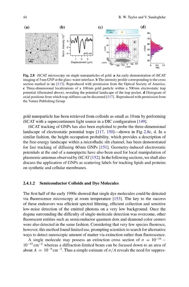
Fig. 2.8 iSCAT microscopy on single nanoparticles of gold. a An early demonstration of iSCAT
imaging of 5 nm GNP at the glass–water interface. b The intensity profile corresponding to the cross
section marked in (a) [113]. Reproduced with permission from the Optical Society of America.
c Three-dimensional localizations of a 100 nm gold particle within a 500 nm electrostatic trap
potential (illustrated above), revealing the potential landscape of the trap pocket. d Histogram of
axial positions from which trap stiffness can be discerned [117]. Reproduced with permission from
the Nature Publishing Group
gold nanoparticle has been retrieved from colloids as small as 10 nm by performing
iSCAT with a supercontinuum light source in a DIC configuration [149].
iSCAT tracking of GNPs has also been exploited to probe the three-dimensional
landscape of electrostatic potential traps [117, 150]—shown in Fig. 2.8c, d. In a
similar fashion, the height occupation probability, which provides a description of
the free-energy landscape within a microfludic slit channel, has been demonstrated
for fast tracking of diffusing 60 nm GNPs [151]. Geometry-induced electrostatic
potentials at the end of a nanopipette have also been used for local manipulation of
plasmonic antennas observed by iSCAT [152]. In the following sections, we shall also
discuss the application of GNPs as scattering labels for tracking lipids and proteins
on synthetic and cellular membranes.
2.4.1.2 Semiconductor Colloids and Dye Molecules
The first half of the early 1990s showed that single dye molecules could be detected
via fluorescence microscopy at room temperature [153]. The key to the success
of these endeavors was efficient spectral filtering, efficient collection and sensitive
low-noise detection of the emitted photons on a very low background. Once the
dogma surrounding the difficulty of single-molecule detection was overcome, other
fluorescent entities such as semiconductor quantum dots and diamond color centers
were also detected in the same fashion. Considering that very few species fluoresce,
however, this method found limited use, prompting scientists to search for alternative
ways to detect nanoscopic amount of matter via extinction rather than fluorescence.
A single molecule may possess an extinction cross section of σ = 10
−16
−
10
−15 cm
−2 whereas a diffraction-limited beam can be focused down to an area of
about A = 10
−9 cm
−2 . Thus a simple estimate of σ/A reveals the need for suppres-
R. W. Taylor and V. Sandoghdar
(a)
(b)
(c)
(d)
electrostatic
trap
Fig. 2.8 iSCAT microscopy on single nanoparticles of gold. a An early demonstration of iSCAT
imaging of 5 nm GNP at the glass–water interface. b The intensity profile corresponding to the cross
section marked in (a) [113]. Reproduced with permission from the Optical Society of America.
c Three-dimensional localizations of a 100 nm gold particle within a 500 nm electrostatic trap
potential (illustrated above), revealing the potential landscape of the trap pocket. d Histogram of
axial positions from which trap stiffness can be discerned [117]. Reproduced with permission from
the Nature Publishing Group
gold nanoparticle has been retrieved from colloids as small as 10 nm by performing
iSCAT with a supercontinuum light source in a DIC configuration [149].
iSCAT tracking of GNPs has also been exploited to probe the three-dimensional
landscape of electrostatic potential traps [117, 150]—shown in Fig. 2.8c, d. In a
similar fashion, the height occupation probability, which provides a description of
the free-energy landscape within a microfludic slit channel, has been demonstrated
for fast tracking of diffusing 60 nm GNPs [151]. Geometry-induced electrostatic
potentials at the end of a nanopipette have also been used for local manipulation of
plasmonic antennas observed by iSCAT [152]. In the following sections, we shall also
discuss the application of GNPs as scattering labels for tracking lipids and proteins
on synthetic and cellular membranes.
2.4.1.2 Semiconductor Colloids and Dye Molecules
The first half of the early 1990s showed that single dye molecules could be detected
via fluorescence microscopy at room temperature [153]. The key to the success
of these endeavors was efficient spectral filtering, efficient collection and sensitive
low-noise detection of the emitted photons on a very low background. Once the
dogma surrounding the difficulty of single-molecule detection was overcome, other
fluorescent entities such as semiconductor quantum dots and diamond color centers
were also detected in the same fashion. Considering that very few species fluoresce,
however, this method found limited use, prompting scientists to search for alternative
ways to detect nanoscopic amount of matter via extinction rather than fluorescence.
A single molecule may possess an extinction cross section of σ = 10
−16
−
10
−15 cm
−2 whereas a diffraction-limited beam can be focused down to an area of
about A = 10
−9 cm
−2 . Thus a simple estimate of σ/A reveals the need for suppres-