

2 Interferometric Scattering (iSCAT) Microscopy and Related Techniques
41
200 nm
2000 nm
20 00 nm
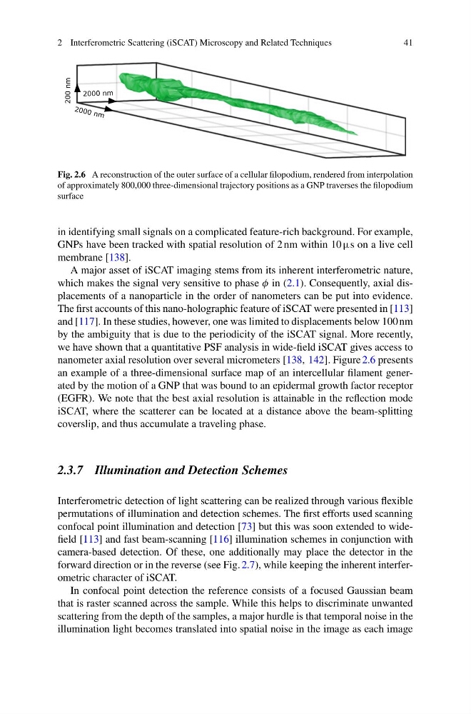
Fig. 2.6 A reconstruction of the outer surface of a cellular filopodium, rendered from interpolation
of approximately 800,000 three-dimensional trajectory positions as a GNP traverses the filopodium
surface
in identifying small signals on a complicated feature-rich background. For example,
GNPs have been tracked with spatial resolution of 2 nm within 10 µs on a live cell
membrane [138].
A major asset of iSCAT imaging stems from its inherent interferometric nature,
which makes the signal very sensitive to phase φ in (2.1). Consequently, axial displacements of a nanoparticle in the order of nanometers can be put into evidence.
The first accounts of this nano-holographic feature of iSCAT were presented in [113]
and [117]. In these studies, however, one was limited to displacements below 100 nm
by the ambiguity that is due to the periodicity of the iSCAT signal. More recently,
we have shown that a quantitative PSF analysis in wide-field iSCAT gives access to
nanometer axial resolution over several micrometers [138, 142]. Figure 2.6 presents
an example of a three-dimensional surface map of an intercellular filament generated by the motion of a GNP that was bound to an epidermal growth factor receptor
(EGFR). We note that the best axial resolution is attainable in the reflection mode
iSCAT, where the scatterer can be located at a distance above the beam-splitting
coverslip, and thus accumulate a traveling phase.
2.3.7 Illumination and Detection Schemes
Interferometric detection of light scattering can be realized through various flexible
permutations of illumination and detection schemes. The first efforts used scanning
confocal point illumination and detection [73] but this was soon extended to widefield [113] and fast beam-scanning [116] illumination schemes in conjunction with
camera-based detection. Of these, one additionally may place the detector in the
forward direction or in the reverse (see Fig. 2.7), while keeping the inherent interferometric character of iSCAT.
In confocal point detection the reference consists of a focused Gaussian beam
that is raster scanned across the sample. While this helps to discriminate unwanted
scattering from the depth of the samples, a major hurdle is that temporal noise in the
illumination light becomes translated into spatial noise in the image as each image
41
200 nm
2000 nm
20 00 nm
Fig. 2.6 A reconstruction of the outer surface of a cellular filopodium, rendered from interpolation
of approximately 800,000 three-dimensional trajectory positions as a GNP traverses the filopodium
surface
in identifying small signals on a complicated feature-rich background. For example,
GNPs have been tracked with spatial resolution of 2 nm within 10 µs on a live cell
membrane [138].
A major asset of iSCAT imaging stems from its inherent interferometric nature,
which makes the signal very sensitive to phase φ in (2.1). Consequently, axial displacements of a nanoparticle in the order of nanometers can be put into evidence.
The first accounts of this nano-holographic feature of iSCAT were presented in [113]
and [117]. In these studies, however, one was limited to displacements below 100 nm
by the ambiguity that is due to the periodicity of the iSCAT signal. More recently,
we have shown that a quantitative PSF analysis in wide-field iSCAT gives access to
nanometer axial resolution over several micrometers [138, 142]. Figure 2.6 presents
an example of a three-dimensional surface map of an intercellular filament generated by the motion of a GNP that was bound to an epidermal growth factor receptor
(EGFR). We note that the best axial resolution is attainable in the reflection mode
iSCAT, where the scatterer can be located at a distance above the beam-splitting
coverslip, and thus accumulate a traveling phase.
2.3.7 Illumination and Detection Schemes
Interferometric detection of light scattering can be realized through various flexible
permutations of illumination and detection schemes. The first efforts used scanning
confocal point illumination and detection [73] but this was soon extended to widefield [113] and fast beam-scanning [116] illumination schemes in conjunction with
camera-based detection. Of these, one additionally may place the detector in the
forward direction or in the reverse (see Fig. 2.7), while keeping the inherent interferometric character of iSCAT.
In confocal point detection the reference consists of a focused Gaussian beam
that is raster scanned across the sample. While this helps to discriminate unwanted
scattering from the depth of the samples, a major hurdle is that temporal noise in the
illumination light becomes translated into spatial noise in the image as each image