

17 Microsphere-Assisted Interference Microscopy
461
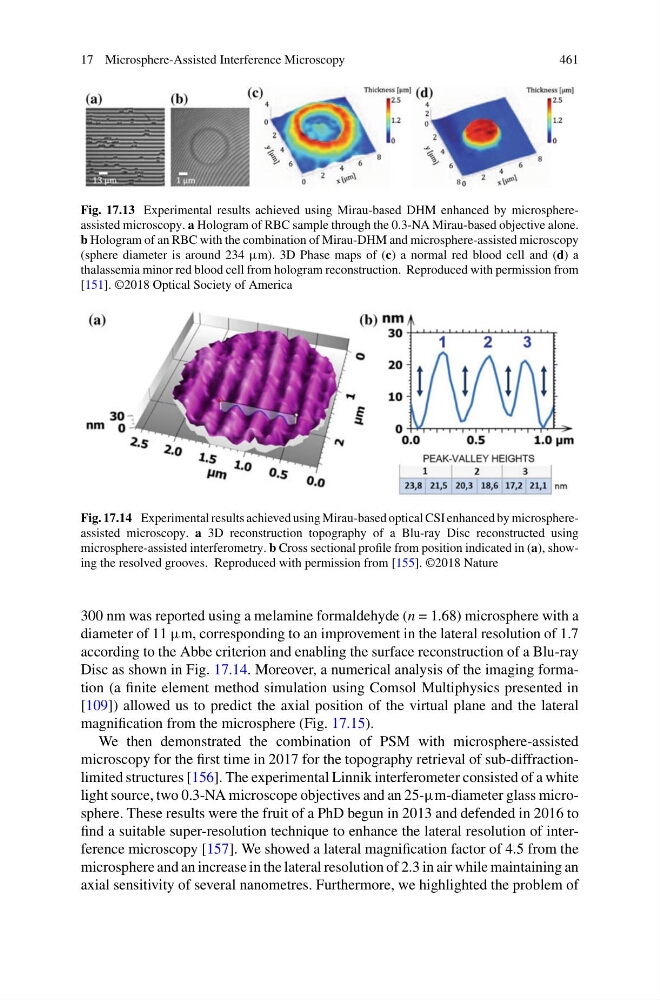
Fig. 17.13 Experimental results achieved using Mirau-based DHM enhanced by microsphereassisted microscopy. a Hologram of RBC sample through the 0.3-NA Mirau-based objective alone.
b Hologram of an RBC with the combination of Mirau-DHM and microsphere-assisted microscopy
(sphere diameter is around 234 µm). 3D Phase maps of (c) a normal red blood cell and (d) a
thalassemia minor red blood cell from hologram reconstruction. Reproduced with permission from
[151]. ©2018 Optical Society of America
Fig. 17.14 Experimental results achieved using Mirau-based optical CSI enhanced by microsphereassisted microscopy. a 3D reconstruction topography of a Blu-ray Disc reconstructed using
microsphere-assisted interferometry. b Cross sectional profile from position indicated in (a), showing the resolved grooves. Reproduced with permission from [155]. ©2018 Nature
300 nm was reported using a melamine formaldehyde (n = 1.68) microsphere with a
diameter of 11 µm, corresponding to an improvement in the lateral resolution of 1.7
according to the Abbe criterion and enabling the surface reconstruction of a Blu-ray
Disc as shown in Fig. 17.14. Moreover, a numerical analysis of the imaging formation (a finite element method simulation using Comsol Multiphysics presented in
[109]) allowed us to predict the axial position of the virtual plane and the lateral
magnification from the microsphere (Fig. 17.15).
We then demonstrated the combination of PSM with microsphere-assisted
microscopy for the first time in 2017 for the topography retrieval of sub-diffractionlimited structures [156]. The experimental Linnik interferometer consisted of a white
light source, two 0.3-NA microscope objectives and an 25-µm-diameter glass microsphere. These results were the fruit of a PhD begun in 2013 and defended in 2016 to
find a suitable super-resolution technique to enhance the lateral resolution of interference microscopy [157]. We showed a lateral magnification factor of 4.5 from the
microsphere and an increase in the lateral resolution of 2.3 in air while maintaining an
axial sensitivity of several nanometres. Furthermore, we highlighted the problem of
461
Fig. 17.13 Experimental results achieved using Mirau-based DHM enhanced by microsphereassisted microscopy. a Hologram of RBC sample through the 0.3-NA Mirau-based objective alone.
b Hologram of an RBC with the combination of Mirau-DHM and microsphere-assisted microscopy
(sphere diameter is around 234 µm). 3D Phase maps of (c) a normal red blood cell and (d) a
thalassemia minor red blood cell from hologram reconstruction. Reproduced with permission from
[151]. ©2018 Optical Society of America
Fig. 17.14 Experimental results achieved using Mirau-based optical CSI enhanced by microsphereassisted microscopy. a 3D reconstruction topography of a Blu-ray Disc reconstructed using
microsphere-assisted interferometry. b Cross sectional profile from position indicated in (a), showing the resolved grooves. Reproduced with permission from [155]. ©2018 Nature
300 nm was reported using a melamine formaldehyde (n = 1.68) microsphere with a
diameter of 11 µm, corresponding to an improvement in the lateral resolution of 1.7
according to the Abbe criterion and enabling the surface reconstruction of a Blu-ray
Disc as shown in Fig. 17.14. Moreover, a numerical analysis of the imaging formation (a finite element method simulation using Comsol Multiphysics presented in
[109]) allowed us to predict the axial position of the virtual plane and the lateral
magnification from the microsphere (Fig. 17.15).
We then demonstrated the combination of PSM with microsphere-assisted
microscopy for the first time in 2017 for the topography retrieval of sub-diffractionlimited structures [156]. The experimental Linnik interferometer consisted of a white
light source, two 0.3-NA microscope objectives and an 25-µm-diameter glass microsphere. These results were the fruit of a PhD begun in 2013 and defended in 2016 to
find a suitable super-resolution technique to enhance the lateral resolution of interference microscopy [157]. We showed a lateral magnification factor of 4.5 from the
microsphere and an increase in the lateral resolution of 2.3 in air while maintaining an
axial sensitivity of several nanometres. Furthermore, we highlighted the problem of