

462
S. Perrin et al.
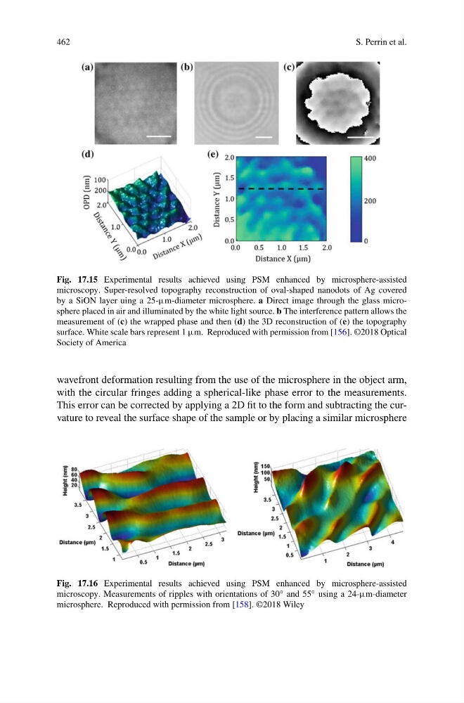
Fig. 17.15 Experimental results achieved using PSM enhanced by microsphere-assisted
microscopy. Super-resolved topography reconstruction of oval-shaped nanodots of Ag covered
by a SiON layer uing a 25-µm-diameter microsphere. a Direct image through the glass microsphere placed in air and illuminated by the white light source. b The interference pattern allows the
measurement of (c) the wrapped phase and then (d) the 3D reconstruction of (e) the topography
surface. White scale bars represent 1 µm. Reproduced with permission from [156]. ©2018 Optical
Society of America
wavefront deformation resulting from the use of the microsphere in the object arm,
with the circular fringes adding a spherical-like phase error to the measurements.
This error can be corrected by applying a 2D fit to the form and subtracting the curvature to reveal the surface shape of the sample or by placing a similar microsphere
Fig. 17.16 Experimental results achieved using PSM enhanced by microsphere-assisted
microscopy. Measurements of ripples with orientations of 30 ◦ and 55 ◦ using a 24-µm-diameter
microsphere. Reproduced with permission from [158]. ©2018 Wiley
S. Perrin et al.
Fig. 17.15 Experimental results achieved using PSM enhanced by microsphere-assisted
microscopy. Super-resolved topography reconstruction of oval-shaped nanodots of Ag covered
by a SiON layer uing a 25-µm-diameter microsphere. a Direct image through the glass microsphere placed in air and illuminated by the white light source. b The interference pattern allows the
measurement of (c) the wrapped phase and then (d) the 3D reconstruction of (e) the topography
surface. White scale bars represent 1 µm. Reproduced with permission from [156]. ©2018 Optical
Society of America
wavefront deformation resulting from the use of the microsphere in the object arm,
with the circular fringes adding a spherical-like phase error to the measurements.
This error can be corrected by applying a 2D fit to the form and subtracting the curvature to reveal the surface shape of the sample or by placing a similar microsphere
Fig. 17.16 Experimental results achieved using PSM enhanced by microsphere-assisted
microscopy. Measurements of ripples with orientations of 30 ◦ and 55 ◦ using a 24-µm-diameter
microsphere. Reproduced with permission from [158]. ©2018 Wiley