

460
S. Perrin et al.
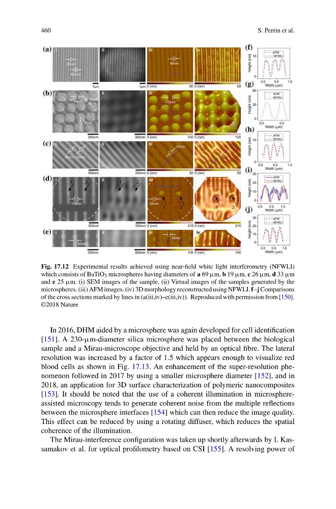
Fig. 17.12 Experimental results achieved using near-field white light interferometry (NFWLI)
which consists of BaTiO 3 microspheres having diameters of a 69 µm, b 19 µm, c 26 µm, d 33 µm
and e 25 µm. (i) SEM images of the sample. (ii) Virtual images of the samples generated by the
microspheres. (iii) AFM images. (iv) 3D morphology reconstructed using NFWLI. f–j Comparisons
of the cross sections marked by lines in (a(iii,iv)–e(iii,iv)). Reproduced with permission from [150].
©2018 Nature
In 2016, DHM aided by a microsphere was again developed for cell identification
[151]. A 230-µm-diameter silica microsphere was placed between the biological
sample and a Mirau-microscope objective and held by an optical fibre. The lateral
resolution was increased by a factor of 1.5 which appears enough to visualize red
blood cells as shown in Fig. 17.13. An enhancement of the super-resolution phenomenon followed in 2017 by using a smaller microsphere diameter [152], and in
2018, an application for 3D surface characterization of polymeric nanocomposites
[153]. It should be noted that the use of a coherent illumination in microsphereassisted microscopy tends to generate coherent noise from the multiple reflections
between the microsphere interfaces [154] which can then reduce the image quality.
This effect can be reduced by using a rotating diffuser, which reduces the spatial
coherence of the illumination.
The Mirau-interference configuration was taken up shortly afterwards by I. Kassamakov et al. for optical profilometry based on CSI [155]. A resolving power of
S. Perrin et al.
Fig. 17.12 Experimental results achieved using near-field white light interferometry (NFWLI)
which consists of BaTiO 3 microspheres having diameters of a 69 µm, b 19 µm, c 26 µm, d 33 µm
and e 25 µm. (i) SEM images of the sample. (ii) Virtual images of the samples generated by the
microspheres. (iii) AFM images. (iv) 3D morphology reconstructed using NFWLI. f–j Comparisons
of the cross sections marked by lines in (a(iii,iv)–e(iii,iv)). Reproduced with permission from [150].
©2018 Nature
In 2016, DHM aided by a microsphere was again developed for cell identification
[151]. A 230-µm-diameter silica microsphere was placed between the biological
sample and a Mirau-microscope objective and held by an optical fibre. The lateral
resolution was increased by a factor of 1.5 which appears enough to visualize red
blood cells as shown in Fig. 17.13. An enhancement of the super-resolution phenomenon followed in 2017 by using a smaller microsphere diameter [152], and in
2018, an application for 3D surface characterization of polymeric nanocomposites
[153]. It should be noted that the use of a coherent illumination in microsphereassisted microscopy tends to generate coherent noise from the multiple reflections
between the microsphere interfaces [154] which can then reduce the image quality.
This effect can be reduced by using a rotating diffuser, which reduces the spatial
coherence of the illumination.
The Mirau-interference configuration was taken up shortly afterwards by I. Kassamakov et al. for optical profilometry based on CSI [155]. A resolving power of