

454
S. Perrin et al.
(c.ii.)
(c.i.)
(b.ii.)
(b.i.)
(a.ii.)
(a.i.)
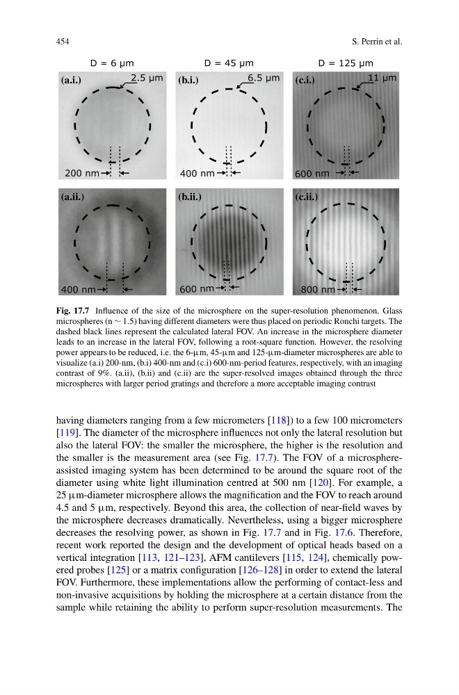
Fig. 17.7 Influence of the size of the microsphere on the super-resolution phenomenon. Glass
microspheres (n ∼ 1.5) having different diameters were thus placed on periodic Ronchi targets. The
dashed black lines represent the calculated lateral FOV. An increase in the microsphere diameter
leads to an increase in the lateral FOV, following a root-square function. However, the resolving
power appears to be reduced, i.e. the 6-µm, 45-µm and 125-µm-diameter microspheres are able to
visualize (a.i) 200-nm, (b.i) 400-nm and (c.i) 600-nm-period features, respectively, with an imaging
contrast of 9%. (a.ii), (b.ii) and (c.ii) are the super-resolved images obtained through the three
microspheres with larger period gratings and therefore a more acceptable imaging contrast
having diameters ranging from a few micrometers [118]) to a few 100 micrometers
[119]. The diameter of the microsphere influences not only the lateral resolution but
also the lateral FOV: the smaller the microsphere, the higher is the resolution and
the smaller is the measurement area (see Fig. 17.7). The FOV of a microsphereassisted imaging system has been determined to be around the square root of the
diameter using white light illumination centred at 500 nm [120]. For example, a
25 µm-diameter microsphere allows the magnification and the FOV to reach around
4.5 and 5 µm, respectively. Beyond this area, the collection of near-field waves by
the microsphere decreases dramatically. Nevertheless, using a bigger microsphere
decreases the resolving power, as shown in Fig. 17.7 and in Fig. 17.6. Therefore,
recent work reported the design and the development of optical heads based on a
vertical integration [113, 121–123], AFM cantilevers [115, 124], chemically powered probes [125] or a matrix configuration [126–128] in order to extend the lateral
FOV. Furthermore, these implementations allow the performing of contact-less and
non-invasive acquisitions by holding the microsphere at a certain distance from the
sample while retaining the ability to perform super-resolution measurements. The
S. Perrin et al.
(c.ii.)
(c.i.)
(b.ii.)
(b.i.)
(a.ii.)
(a.i.)
Fig. 17.7 Influence of the size of the microsphere on the super-resolution phenomenon. Glass
microspheres (n ∼ 1.5) having different diameters were thus placed on periodic Ronchi targets. The
dashed black lines represent the calculated lateral FOV. An increase in the microsphere diameter
leads to an increase in the lateral FOV, following a root-square function. However, the resolving
power appears to be reduced, i.e. the 6-µm, 45-µm and 125-µm-diameter microspheres are able to
visualize (a.i) 200-nm, (b.i) 400-nm and (c.i) 600-nm-period features, respectively, with an imaging
contrast of 9%. (a.ii), (b.ii) and (c.ii) are the super-resolved images obtained through the three
microspheres with larger period gratings and therefore a more acceptable imaging contrast
having diameters ranging from a few micrometers [118]) to a few 100 micrometers
[119]. The diameter of the microsphere influences not only the lateral resolution but
also the lateral FOV: the smaller the microsphere, the higher is the resolution and
the smaller is the measurement area (see Fig. 17.7). The FOV of a microsphereassisted imaging system has been determined to be around the square root of the
diameter using white light illumination centred at 500 nm [120]. For example, a
25 µm-diameter microsphere allows the magnification and the FOV to reach around
4.5 and 5 µm, respectively. Beyond this area, the collection of near-field waves by
the microsphere decreases dramatically. Nevertheless, using a bigger microsphere
decreases the resolving power, as shown in Fig. 17.7 and in Fig. 17.6. Therefore,
recent work reported the design and the development of optical heads based on a
vertical integration [113, 121–123], AFM cantilevers [115, 124], chemically powered probes [125] or a matrix configuration [126–128] in order to extend the lateral
FOV. Furthermore, these implementations allow the performing of contact-less and
non-invasive acquisitions by holding the microsphere at a certain distance from the
sample while retaining the ability to perform super-resolution measurements. The