

17 Microsphere-Assisted Interference Microscopy
453
Fig. 17.5 Comparison of the
modulus of the contrast
transfer function (CTF) of
microsphere-assisted
microscopy with optical
microscopy. The frequency
response has been measured
using positive Ronchi
targets. The microsphere has
a refractive index of 1.52 and
a diameter of 26 µm. The
illumination is a white light
source [116]
1.0
0.8
0.6
0.4
0.2
Modulus of the CTF (a.u.)
0.0
0.5 1.0 1.5 2.0 2.5 3.0
5.0
Spatial frequency (cycles/μm)
3.5 4.0 4.5
Microsphere-assisted microscopy
Optical microscopy (x50/0.55 objective)
0.0
CTF = 27%
CTF = 22%
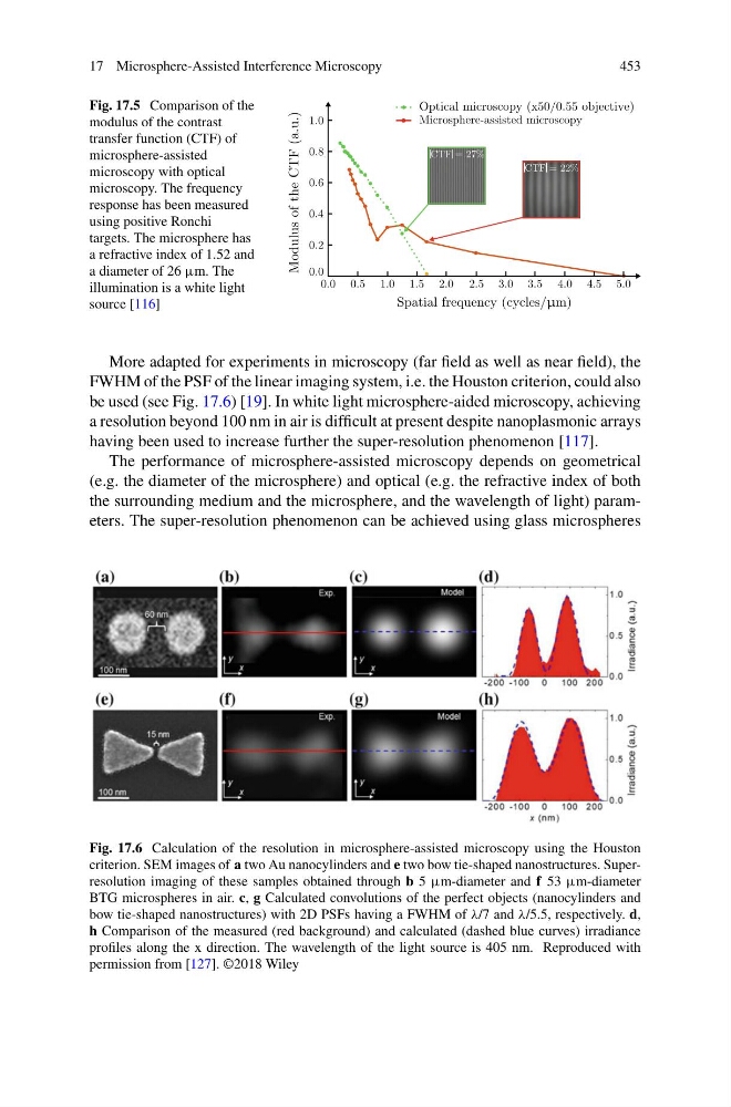
More adapted for experiments in microscopy (far field as well as near field), the
FWHM of the PSF of the linear imaging system, i.e. the Houston criterion, could also
be used (see Fig. 17.6) [19]. In white light microsphere-aided microscopy, achieving
a resolution beyond 100 nm in air is difficult at present despite nanoplasmonic arrays
having been used to increase further the super-resolution phenomenon [117].
The performance of microsphere-assisted microscopy depends on geometrical
(e.g. the diameter of the microsphere) and optical (e.g. the refractive index of both
the surrounding medium and the microsphere, and the wavelength of light) parameters. The super-resolution phenomenon can be achieved using glass microspheres
Fig. 17.6 Calculation of the resolution in microsphere-assisted microscopy using the Houston
criterion. SEM images of a two Au nanocylinders and e two bow tie-shaped nanostructures. Superresolution imaging of these samples obtained through b 5 µm-diameter and f 53 µm-diameter
BTG microspheres in air. c, g Calculated convolutions of the perfect objects (nanocylinders and
bow tie-shaped nanostructures) with 2D PSFs having a FWHM of λ/7 and λ/5.5, respectively. d,
h Comparison of the measured (red background) and calculated (dashed blue curves) irradiance
profiles along the x direction. The wavelength of the light source is 405 nm. Reproduced with
permission from [127]. ©2018 Wiley
453
Fig. 17.5 Comparison of the
modulus of the contrast
transfer function (CTF) of
microsphere-assisted
microscopy with optical
microscopy. The frequency
response has been measured
using positive Ronchi
targets. The microsphere has
a refractive index of 1.52 and
a diameter of 26 µm. The
illumination is a white light
source [116]
1.0
0.8
0.6
0.4
0.2
Modulus of the CTF (a.u.)
0.0
0.5 1.0 1.5 2.0 2.5 3.0
5.0
Spatial frequency (cycles/μm)
3.5 4.0 4.5
Microsphere-assisted microscopy
Optical microscopy (x50/0.55 objective)
0.0
CTF = 27%
CTF = 22%
More adapted for experiments in microscopy (far field as well as near field), the
FWHM of the PSF of the linear imaging system, i.e. the Houston criterion, could also
be used (see Fig. 17.6) [19]. In white light microsphere-aided microscopy, achieving
a resolution beyond 100 nm in air is difficult at present despite nanoplasmonic arrays
having been used to increase further the super-resolution phenomenon [117].
The performance of microsphere-assisted microscopy depends on geometrical
(e.g. the diameter of the microsphere) and optical (e.g. the refractive index of both
the surrounding medium and the microsphere, and the wavelength of light) parameters. The super-resolution phenomenon can be achieved using glass microspheres
Fig. 17.6 Calculation of the resolution in microsphere-assisted microscopy using the Houston
criterion. SEM images of a two Au nanocylinders and e two bow tie-shaped nanostructures. Superresolution imaging of these samples obtained through b 5 µm-diameter and f 53 µm-diameter
BTG microspheres in air. c, g Calculated convolutions of the perfect objects (nanocylinders and
bow tie-shaped nanostructures) with 2D PSFs having a FWHM of λ/7 and λ/5.5, respectively. d,
h Comparison of the measured (red background) and calculated (dashed blue curves) irradiance
profiles along the x direction. The wavelength of the light source is 405 nm. Reproduced with
permission from [127]. ©2018 Wiley