

17 Microsphere-Assisted Interference Microscopy
447
Optical Nanoscopy
Perfect lens
Hyperbolic lens
Microsphere
Resolved
Detected
Full field
Scanning
Far field
Near field
SNOM / PSTM
FRET
LST
SEEC
TSOM
CSM
4Pi
SIM
TIRF
PSM
CSI
DHM
TDM
STED
Axial
Lateral
Axial
Lateral
PALM
STORM
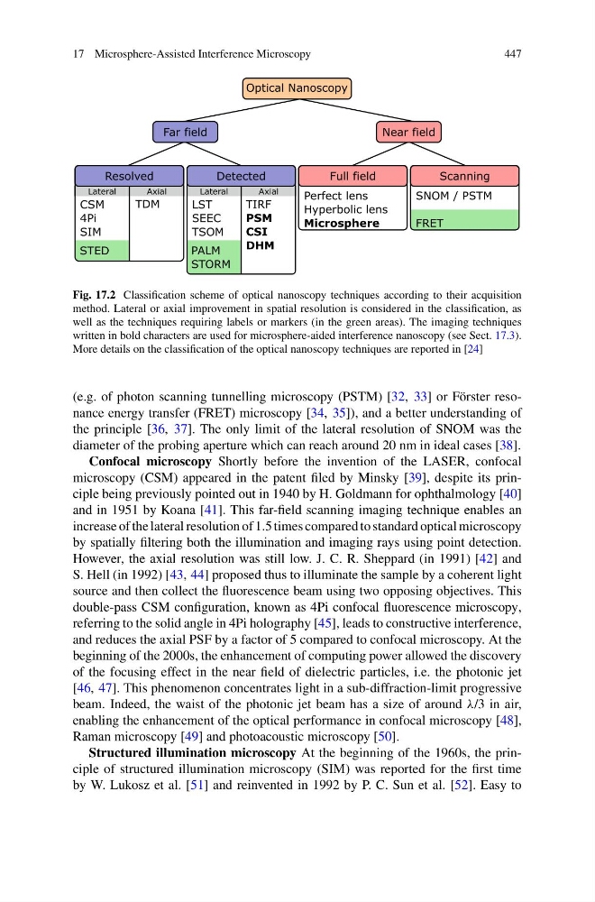
Fig. 17.2 Classification scheme of optical nanoscopy techniques according to their acquisition
method. Lateral or axial improvement in spatial resolution is considered in the classification, as
well as the techniques requiring labels or markers (in the green areas). The imaging techniques
written in bold characters are used for microsphere-aided interference nanoscopy (see Sect. 17.3).
More details on the classification of the optical nanoscopy techniques are reported in [24]
(e.g. of photon scanning tunnelling microscopy (PSTM) [32, 33] or Förster resonance energy transfer (FRET) microscopy [34, 35]), and a better understanding of
the principle [36, 37]. The only limit of the lateral resolution of SNOM was the
diameter of the probing aperture which can reach around 20 nm in ideal cases [38].
Confocal microscopy Shortly before the invention of the LASER, confocal
microscopy (CSM) appeared in the patent filed by Minsky [39], despite its principle being previously pointed out in 1940 by H. Goldmann for ophthalmology [40]
and in 1951 by Koana [41]. This far-field scanning imaging technique enables an
increase of the lateral resolution of 1.5 times compared to standard optical microscopy
by spatially filtering both the illumination and imaging rays using point detection.
However, the axial resolution was still low. J. C. R. Sheppard (in 1991) [42] and
S. Hell (in 1992) [43, 44] proposed thus to illuminate the sample by a coherent light
source and then collect the fluorescence beam using two opposing objectives. This
double-pass CSM configuration, known as 4Pi confocal fluorescence microscopy,
referring to the solid angle in 4Pi holography [45], leads to constructive interference,
and reduces the axial PSF by a factor of 5 compared to confocal microscopy. At the
beginning of the 2000s, the enhancement of computing power allowed the discovery
of the focusing effect in the near field of dielectric particles, i.e. the photonic jet
[46, 47]. This phenomenon concentrates light in a sub-diffraction-limit progressive
beam. Indeed, the waist of the photonic jet beam has a size of around λ/3 in air,
enabling the enhancement of the optical performance in confocal microscopy [48],
Raman microscopy [49] and photoacoustic microscopy [50].
Structured illumination microscopy At the beginning of the 1960s, the principle of structured illumination microscopy (SIM) was reported for the first time
by W. Lukosz et al. [51] and reinvented in 1992 by P. C. Sun et al. [52]. Easy to
447
Optical Nanoscopy
Perfect lens
Hyperbolic lens
Microsphere
Resolved
Detected
Full field
Scanning
Far field
Near field
SNOM / PSTM
FRET
LST
SEEC
TSOM
CSM
4Pi
SIM
TIRF
PSM
CSI
DHM
TDM
STED
Axial
Lateral
Axial
Lateral
PALM
STORM
Fig. 17.2 Classification scheme of optical nanoscopy techniques according to their acquisition
method. Lateral or axial improvement in spatial resolution is considered in the classification, as
well as the techniques requiring labels or markers (in the green areas). The imaging techniques
written in bold characters are used for microsphere-aided interference nanoscopy (see Sect. 17.3).
More details on the classification of the optical nanoscopy techniques are reported in [24]
(e.g. of photon scanning tunnelling microscopy (PSTM) [32, 33] or Förster resonance energy transfer (FRET) microscopy [34, 35]), and a better understanding of
the principle [36, 37]. The only limit of the lateral resolution of SNOM was the
diameter of the probing aperture which can reach around 20 nm in ideal cases [38].
Confocal microscopy Shortly before the invention of the LASER, confocal
microscopy (CSM) appeared in the patent filed by Minsky [39], despite its principle being previously pointed out in 1940 by H. Goldmann for ophthalmology [40]
and in 1951 by Koana [41]. This far-field scanning imaging technique enables an
increase of the lateral resolution of 1.5 times compared to standard optical microscopy
by spatially filtering both the illumination and imaging rays using point detection.
However, the axial resolution was still low. J. C. R. Sheppard (in 1991) [42] and
S. Hell (in 1992) [43, 44] proposed thus to illuminate the sample by a coherent light
source and then collect the fluorescence beam using two opposing objectives. This
double-pass CSM configuration, known as 4Pi confocal fluorescence microscopy,
referring to the solid angle in 4Pi holography [45], leads to constructive interference,
and reduces the axial PSF by a factor of 5 compared to confocal microscopy. At the
beginning of the 2000s, the enhancement of computing power allowed the discovery
of the focusing effect in the near field of dielectric particles, i.e. the photonic jet
[46, 47]. This phenomenon concentrates light in a sub-diffraction-limit progressive
beam. Indeed, the waist of the photonic jet beam has a size of around λ/3 in air,
enabling the enhancement of the optical performance in confocal microscopy [48],
Raman microscopy [49] and photoacoustic microscopy [50].
Structured illumination microscopy At the beginning of the 1960s, the principle of structured illumination microscopy (SIM) was reported for the first time
by W. Lukosz et al. [51] and reinvented in 1992 by P. C. Sun et al. [52]. Easy to