

446
S. Perrin et al.
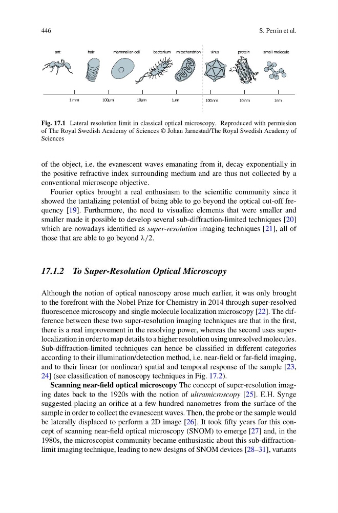
Fig. 17.1 Lateral resolution limit in classical optical microscopy. Reproduced with permission
of The Royal Swedish Academy of Sciences © Johan Jarnestad/The Royal Swedish Academy of
Sciences
of the object, i.e. the evanescent waves emanating from it, decay exponentially in
the positive refractive index surrounding medium and are thus not collected by a
conventional microscope objective.
Fourier optics brought a real enthusiasm to the scientific community since it
showed the tantalizing potential of being able to go beyond the optical cut-off frequency [19]. Furthermore, the need to visualize elements that were smaller and
smaller made it possible to develop several sub-diffraction-limited techniques [20]
which are nowadays identified as super-resolution imaging techniques [21], all of
those that are able to go beyond λ/2.
17.1.2 To Super-Resolution Optical Microscopy
Although the notion of optical nanoscopy arose much earlier, it was only brought
to the forefront with the Nobel Prize for Chemistry in 2014 through super-resolved
fluorescence microscopy and single molecule localization microscopy [22]. The difference between these two super-resolution imaging techniques are that in the first,
there is a real improvement in the resolving power, whereas the second uses superlocalization in order to map details to a higher resolution using unresolved molecules.
Sub-diffraction-limited techniques can hence be classified in different categories
according to their illumination/detection method, i.e. near-field or far-field imaging,
and to their linear (or nonlinear) spatial and temporal response of the sample [23,
24] (see classification of nanoscopy techniques in Fig. 17.2).
Scanning near-field optical microscopy The concept of super-resolution imaging dates back to the 1920s with the notion of ultramicroscopy [25]. E.H. Synge
suggested placing an orifice at a few hundred nanometres from the surface of the
sample in order to collect the evanescent waves. Then, the probe or the sample would
be laterally displaced to perform a 2D image [26]. It took fifty years for this concept of scanning near-field optical microscopy (SNOM) to emerge [27] and, in the
1980s, the microscopist community became enthusiastic about this sub-diffractionlimit imaging technique, leading to new designs of SNOM devices [28–31], variants
S. Perrin et al.
Fig. 17.1 Lateral resolution limit in classical optical microscopy. Reproduced with permission
of The Royal Swedish Academy of Sciences © Johan Jarnestad/The Royal Swedish Academy of
Sciences
of the object, i.e. the evanescent waves emanating from it, decay exponentially in
the positive refractive index surrounding medium and are thus not collected by a
conventional microscope objective.
Fourier optics brought a real enthusiasm to the scientific community since it
showed the tantalizing potential of being able to go beyond the optical cut-off frequency [19]. Furthermore, the need to visualize elements that were smaller and
smaller made it possible to develop several sub-diffraction-limited techniques [20]
which are nowadays identified as super-resolution imaging techniques [21], all of
those that are able to go beyond λ/2.
17.1.2 To Super-Resolution Optical Microscopy
Although the notion of optical nanoscopy arose much earlier, it was only brought
to the forefront with the Nobel Prize for Chemistry in 2014 through super-resolved
fluorescence microscopy and single molecule localization microscopy [22]. The difference between these two super-resolution imaging techniques are that in the first,
there is a real improvement in the resolving power, whereas the second uses superlocalization in order to map details to a higher resolution using unresolved molecules.
Sub-diffraction-limited techniques can hence be classified in different categories
according to their illumination/detection method, i.e. near-field or far-field imaging,
and to their linear (or nonlinear) spatial and temporal response of the sample [23,
24] (see classification of nanoscopy techniques in Fig. 17.2).
Scanning near-field optical microscopy The concept of super-resolution imaging dates back to the 1920s with the notion of ultramicroscopy [25]. E.H. Synge
suggested placing an orifice at a few hundred nanometres from the surface of the
sample in order to collect the evanescent waves. Then, the probe or the sample would
be laterally displaced to perform a 2D image [26]. It took fifty years for this concept of scanning near-field optical microscopy (SNOM) to emerge [27] and, in the
1980s, the microscopist community became enthusiastic about this sub-diffractionlimit imaging technique, leading to new designs of SNOM devices [28–31], variants