

448
S. Perrin et al.
implement, SIM consists of illuminating the sample with a sinusoidal pattern and
recording at least three angle-shifted images. The period of the periodic excitation
pattern is chosen to shift the inaccessible high-frequency components within the
passband of the conventional optical transfer function (OTF). Using the computation optical sensing and imaging (COSI) approach, the high-resolution information
is extracted and the image is then reconstructed using a deconvolution of the sample with the illumination pattern in Fourier space [53]. The first convincing results
with linear SIM have shown a real enhancement of the lateral resolution and an
overcoming of the diffraction limit by a factor of two [54, 55].
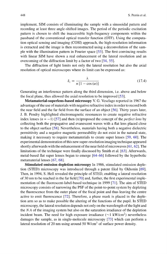
The diffraction of light limits not only the lateral resolution but also the axial
resolution of optical microscopes where its limit can be expressed as:
δ z =
λ
n [1 − cos (α)]
(17.4)
Generating an interference pattern along the third dimension, i.e. above and below
the focal plane, thus allowed the axial resolution to be improved [53].
Metamaterial-superlens-based microsopy V. G. Veselago reported in 1967 the
advantage of the use of materials with negative refractive index in order to record both
the near field and the far field from the surface of an object [56]. Thirty years later,
J. B. Pendry highlighted electromagnetic resonances to create negative refractive
index lenses (n = −1) [57] and then (re)proposed the concept of the perfect lens by
collecting both the propagative and evanescent waves with a flat lens placed close
to the object surface [58]. Nevertheless, materials having both a negative dielectric
permittivity and a negative magnetic permeability do not exist in the natural state,
making it necessary to require metamaterials to create super lenses [59, 60]. The
experimental demonstration of this new super-resolution imaging technique appeared
shortly afterwards with the enhancement of the near field of microwaves [61, 62]. The
limitations of the technique were finally discussed by Smith et al. [63]. Afterwards,
metal-based flat super lenses began to emerge [64–66] followed by the hyperbolic
metamaterial lenses [67, 68].
Stimulated emission depletion microsopy In 1986, stimulated emission depletion (STED) microscopy was introduced through a patent filed by Okhonin [69].
Then, in 1994, S. Hell revealed the principle of STED, enabling a lateral resolution
of 30 nm to be reached in the far field [70] and, further, the first experimental implementation of the fluorescent-label-based technique in 1999 [71]. The aim of STED
microscopy consists of narrowing the PSF of the point-to-point system by depleting
the fluorescence from the outer plane of the focal point and thus leaving the centre
active to emit fluorescence [72]. Therefore, a phase mask is placed in the depletion arm so as to make possible the altering of the functions of the pupil. In STED
microscopy, the lateral resolution depends not only on the wavelength of the light and
the N A of the imaging system but also on the saturation irradiance of the depletion
incident beam. The need for high exposure irradiance (∼1 kW/cm
2 ) nevertheless
damages the sample, as in single-molecule microscopy [73] which can perform a
lateral resolution of 20 nm using around 50 W/cm
2 of surface power density.
S. Perrin et al.
implement, SIM consists of illuminating the sample with a sinusoidal pattern and
recording at least three angle-shifted images. The period of the periodic excitation
pattern is chosen to shift the inaccessible high-frequency components within the
passband of the conventional optical transfer function (OTF). Using the computation optical sensing and imaging (COSI) approach, the high-resolution information
is extracted and the image is then reconstructed using a deconvolution of the sample with the illumination pattern in Fourier space [53]. The first convincing results
with linear SIM have shown a real enhancement of the lateral resolution and an
overcoming of the diffraction limit by a factor of two [54, 55].
The diffraction of light limits not only the lateral resolution but also the axial
resolution of optical microscopes where its limit can be expressed as:
δ z =
λ
n [1 − cos (α)]
(17.4)
Generating an interference pattern along the third dimension, i.e. above and below
the focal plane, thus allowed the axial resolution to be improved [53].
Metamaterial-superlens-based microsopy V. G. Veselago reported in 1967 the
advantage of the use of materials with negative refractive index in order to record both
the near field and the far field from the surface of an object [56]. Thirty years later,
J. B. Pendry highlighted electromagnetic resonances to create negative refractive
index lenses (n = −1) [57] and then (re)proposed the concept of the perfect lens by
collecting both the propagative and evanescent waves with a flat lens placed close
to the object surface [58]. Nevertheless, materials having both a negative dielectric
permittivity and a negative magnetic permeability do not exist in the natural state,
making it necessary to require metamaterials to create super lenses [59, 60]. The
experimental demonstration of this new super-resolution imaging technique appeared
shortly afterwards with the enhancement of the near field of microwaves [61, 62]. The
limitations of the technique were finally discussed by Smith et al. [63]. Afterwards,
metal-based flat super lenses began to emerge [64–66] followed by the hyperbolic
metamaterial lenses [67, 68].
Stimulated emission depletion microsopy In 1986, stimulated emission depletion (STED) microscopy was introduced through a patent filed by Okhonin [69].
Then, in 1994, S. Hell revealed the principle of STED, enabling a lateral resolution
of 30 nm to be reached in the far field [70] and, further, the first experimental implementation of the fluorescent-label-based technique in 1999 [71]. The aim of STED
microscopy consists of narrowing the PSF of the point-to-point system by depleting
the fluorescence from the outer plane of the focal point and thus leaving the centre
active to emit fluorescence [72]. Therefore, a phase mask is placed in the depletion arm so as to make possible the altering of the functions of the pupil. In STED
microscopy, the lateral resolution depends not only on the wavelength of the light and
the N A of the imaging system but also on the saturation irradiance of the depletion
incident beam. The need for high exposure irradiance (∼1 kW/cm
2 ) nevertheless
damages the sample, as in single-molecule microscopy [73] which can perform a
lateral resolution of 20 nm using around 50 W/cm
2 of surface power density.